Molecular Biology
DNA (deoxyribonucleic acid) is a nucleic acid made up of a trio of components called nucleotides. Every cell contains DNA either in its cytoplasm (as in prokaryotes) or inside the nucleus (as in eukaryotes). DNA is the only molecule known that is capable of replicating itself. This characteristic allows DNA to permit cell division, which is necessary for tissue growth or tissue repair
.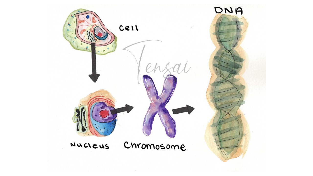
The DNA is located inside the nucleus in eukaryotic cells.
In the 1940s, Biologists began to accept the hypothesis that genetic material was found within chromosomes inside the cell. A chromosome is a long thread of DNA in almost equal amounts with proteins called histones. Prokaryotes have circular chromosomes, while eukaryotes have linear chromosomes. Nucleic Acids are the basic units of a nucleotide. Nucleotides are made up of phosphates, sugar molecules and one of four different nitrogen bases: adenine (A), guanine (G), cytosine (C) & thymine (T).
In 1950, Rosalind Franklin developed a technique of using X-rays to take pictures of the DNA molecule. Later in 1953, James Watson & Francis Crick developed a 3-dimensional model of the DNA molecule. In what would be described as the Watson-Crick model of DNA, the DNA molecule is a double helix resembling a twisted ladder.
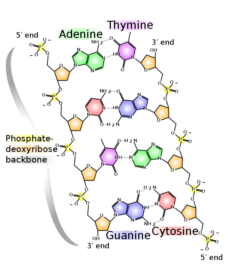
Image: The double helix structure of DNA. (Source: Wikipedia-CC BY-SA 3.0)
The uprights of the DNA 'ladder' are composed of phosphates & deoxyribose sugar. The bases form the “rungs” of the ladder. The sugar and phosphate form the “uprights”. Human DNA is about 3 billion base pairs and about 2 meters in length. The 4 nitrogenous bases can be categorized as purines or pyrimidines. The two purines are adenine and guanine, both with double rings. The two pyrimidines are thymine and cytosine, with single ring structures. Cytosine pairs with guanine- CG (Triple hydrogen bond). Adenine pairs with thymine- AT (Double hydrogen bond). Therefore, a pyrimidine pairs with a purine.
Two adjacent nucleotides are bonded to each other by a Phosphodiester Bond, between the 3' end of the growing strand to the 5' residue of the pentose sugar of the added nucleotide. Because of this bonding nature, the DNA strand has a 3'OH end and a 5'PO4 end. The two strands of DNA that form a double helix are antiparallel, which means the two strands bond in a way so that the 3' end of one DNA is aligned with the 5' end of the complementary strand.
The theory states that Chromosomes carry genes, the units of heredity. Paired chromosomes (homologous chromosomes) segregate during meiosis (gamete formation - Gametogenesis). Each gamete (sex cells - ova/egg or sperm) has half the number of chromosomes found in a somatic (body) cell. In other words, somatic/body cells are diploid (2n) and gametes are Haploid (n).
Chromosomes sort independently during meiosis. Each gamete receives one of the homologous pairs and that one chromosome has no influence on the movement of a member of another pair.
A single chromosome contains many genes linked together and so does the other chromosome in the homologous pair. The sequence of genes on each homologous chromosome pair should match each other exactly. Parts of a chromosome holding many genes, may separate and switch places with the matching part of the other chromosome. This is called crossing over or recombination. The closer genes are to each other, the less likely they will separate during crossing over. These closely located genes are referred to as Linked genes. Gene linkage, therefore, reduces the chance for genetic recombination and variation among offspring
.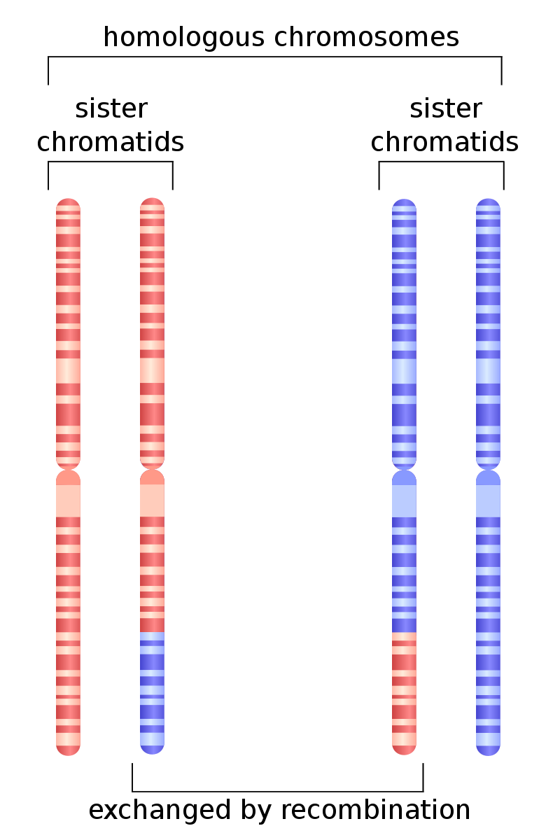
DNA recombination. (Source: Wikipedia-CC BY-SA 3.0)
The Recombination frequencies of genes can be used to determine their positions on chromosomes. This is called Linkage Mapping. recombination frequency (expressed as %) = (number of recombinations * 100) / total number of offspring Gene linkage maps are constructed by ordering fragments of DNA, studying chromosomal alterations and performing crosses to see how frequently crossing over occurs between fragments.
DNA replication is the process of DNA duplicating itself therefore it can also be described as DNA synthesis. DNA replication is necessary in both mitosis and Meiosis.
The Origin of Replication is made up of a particular DNA sequence at which replication is initiated. There are similar characteristics in the origin of replication between various species. The sequences at the origin remain highly conserved. Prokaryotes have one origin of replication on their circular chromosome while eukaryotes have multiple origins of replication along their linear chromosomes.
The enzyme Helicase binds at the ORI and breaks the hydrogen bonds between the nitrogenous bases and results in the unwinding of the DNA double helix into single strands. The region with the unwound DNA appears to form a bubble called the Replication bubble. Eukaryotes have several replication bubbles along the length of their DNA, while prokaryotes will only have one bubble. From the center of the replication bubble, where the unwinding starts, the half side can be described as a Replication fork.
An enzyme called Topoisomerase helps to relieve the stress on the unwinding DNA by causing breaks on the DNA and then resealing the DNA.
To prevent the hydrogen bonds from reforming, the single strand binding proteins (SSBPs) bind at the replication form, stabilizing the DNA strands and preventing them from winding.
This is the pairing of nitrogenous bases. Free-floating nucleotides bind to the unzipped strands of DNA. Each strand acts as a template for the new (daughter) strand. The enzyme DNA polymerase III facilitates the insertion of the nucleotides. DNA synthesis uses the existing (parent) strands as templates, and synthesizes two new complementary daughter strands. This is called The Semi Conservative Model because at the end of the DNA replication process, each DNA molecule will contain one parent strand and one daughter strand.
DNA polymerase III can only add nucleotides in the 5' to 3' direction. (see 'General Introduction' for DNA orientation). Since the daughter DNA will need to be antiparallel, the DNA that acts as the template should be in the 3' to 5' direction. Only one of the two template DNA strands is in the 3' to 5' direction, this strand will be replicated continuously and is named The Leading Strand. The other template DNA strand that is in the 5' to 3' direction will be replicated in fragments called the Okazaki Fragments and is called the the lagging strand.
DNA Polymerase III also requires a primer for it to start adding nucleotides onto it. A primer is a short RNA fragment that is complementary to the DNA template. The primer is synthesized by an enzyme called Primase.
Once the okazaki fragments have been fully synthesized, DNA Polymerase I, which has an exonuclease activity, removes the RNA primers and synthesizes new DNA to fill those gaps. Another enzyme (Ligase) seals the gaps between the okazaki fragments to result in one continuous DNA strand.
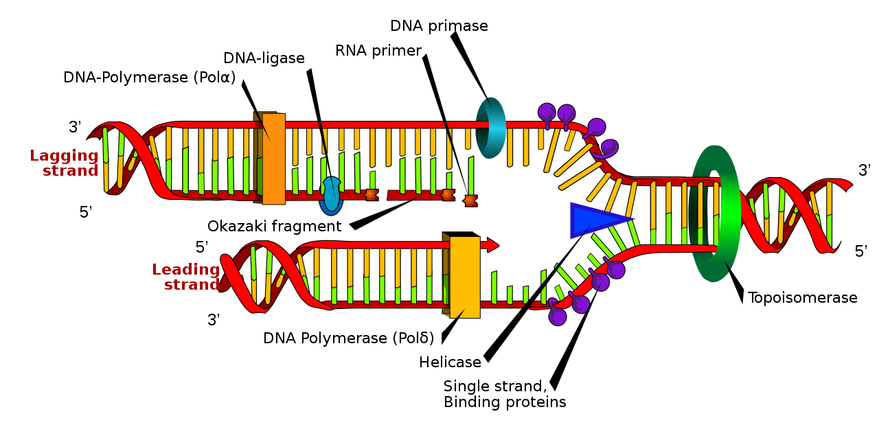
Enzymes involved in DNA replication. (Source: Wikipedia-CC BY-SA 3.0)
DNA replication is highly accurate, however, errors can be corrected through two mechanisms: DNA proofreading and Mismatch repair. DNA Proofreading is performed by the DNA polymerases as they add nucleotides to the daughter strand. The DNA polymerase checks to ensure the correct base was added and that it bonded correctly to the base on the template DNA, before the polymerase moves to the next position. If an incorrect base has been added, the enzyme breaks the phosphodiester bond and releases the incorrect nucleotide and replaces it with a correct matching nucleotide. This can be described as the exonuclease activity of DNA Polymerases. Errors that are not corrected during replication will be corrected through mismatch repair, which occurs at the end of DNA replication. Mismatch repair proteins detect the incorrect base, excises it and replaces it with the correct nucleotide.
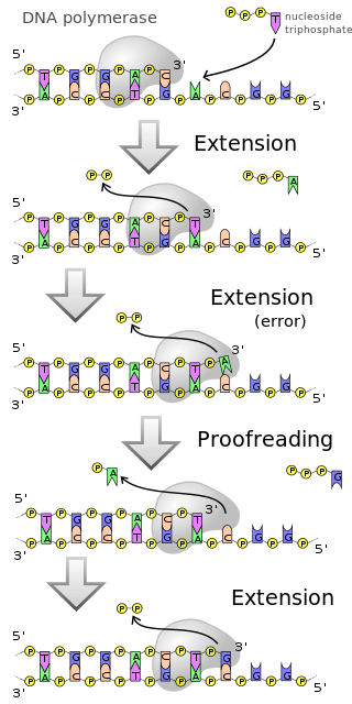
Actions of DNA polymerase. (Source: Wikipedia-CC BY-SA 3.0)
The Central Dogma of Molecular Biology simply describes the flow of information. In this case, it can be summarized as DNA makes RNA (Transcription) and RNA makes Proteins (Translation). Once the information has been transferred to proteins, it can neither be transferred to other proteins, nor back to the RNA or DNA.
DNA Transcription is the synthesis of messenger RNA (mRNA) from DNA. In eukaryotes, it occurs within the nucleus. Transcription can also be defined as Gene expression, therefore transcription occurs when a certain gene is required to be expressed to eventually perform a certain function. The mechanisms of transcription are located in sequences that appear just before and after the gene itself.
DNA transcription process can be broken down into 3 steps.
The initiation of DNA replication occurs at the Promoter site on the DNA strand. Promoter sites are located before the transcription start sites (i.e. upstream). They are about 100-1000 base pairs long. RNA polymerase together with some transcription factors bind to the promoter sequences and creates a transcription bubble, by breaking the hydrogen bonds and separating the two DNA strands. RNA polymerase and the transcription factors select a transcription start site and adds nucleotides complementary to transcription start site.
One strand of DNA is used as the template strand (non-coding strand). RNA polymerase adds free floating nucleotides complementary to the nucleotides on the template strand, with Thymine is replaced with Uracil. RNA synthesis also occurs in the 5' to 3' direction (just like DNA replication), so the template strand needs to be in the 3' to 5' orientation. Elongation also involves proofreading mechanisms that detect and correct mismatched nucleotides. This proofreading process may result in short pauses in the transcription process, especially in eukaryotes.
Termination of transcription occurs when the Transcription termination factor for RNA polymerase encounters the termination sequence. This protein binds at this sequence and causes the RNA polymerase to disengage from the template DNA. (The process of termination is actually more sophisticated than this and learners who want more detailed explanation should review our Cell Biology course).
This describes a set of processes that occur, especially in eukaryotic cells, where the mRNA Primary transcript is modified to form a mature and functional mRNA that can then leave the nucleus and be transported to the cytoplasm to undergo translation. Briefly, these modifications include:
RNA splicing removes introns and links the exons directly, while the 5' cap and 3' tail are necessary for the transport of the mRNA to a ribosome and protect the mRNA from degradation.
Translation (also called Protein Synthesis) is the process by which the mRNA strand is 'translated' into an amino acid sequence (protein). This occurs in the cell organelles called Ribosomes. Ribosomes are located in the cytoplasm as either free floating or fixed onto the rough endoplasmic reticulum. The ribosome type influences what type of proteins it will synthesize.
Mature mRNA is transported from the nucleus into the cytoplasm and it attaches to the respective ribosomes for translation to occur. The ribosome enables the binding of complementary tRNA anticodon sequences to mRNA codons. A codon is set of three nitrogen bases on the mRNA strand that code for one amino acid. An Anticodon is a set of three nitrogen bases located on a tRNA that a complementary to the codon. Not all codons code for amino acids. There are 4 codes that code for initiator or terminator codons. These are codons that tell protein synthesis to start or stop. The tRNAs carry specific amino acids that are chained together into a polypeptide as the mRNA passes through and is read by the ribosome.
A detailed illustration of DNA translation. (Source: Wikipedia-CC BY-SA 3.0)
Translation proceeds in three phases:
Initiation: The ribosome assembles around the mRNA. The first tRNA is attached at the start codon. This start codon always corresponds to a Methionine.
Elongation: The tRNA transfers the corresponding amino acid to the tRNA corresponding to the next codon. The ribosome then moves (translocates) to the next mRNA codon to continue adding an additional amino acid, creating an amino acid chain.
Termination: When a peptidyl tRNA encounters a stop codon, then the ribosome folds the polypeptide into its final structure.
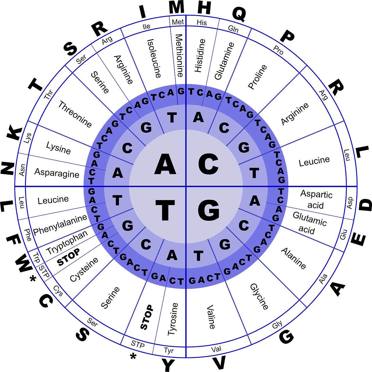
The redundancy of the genetic code.
Mutations are inheritable changes in the genetic (DNA) sequence. Although mutations can arise from mistakes in DNA replication when one nitrogen base is substituted for another, they can also occur outside of DNA replication. Mutations can be caused by mutagenic agents such as X-rays, UV rays, and chemicals that can alter DNA.
Mutations can be categorized in various ways. In one way, mutations can be classified according to the change that occurred on the DNA sequence. In this case there can be Substitutions, Insertions and Deletions. Mutations can also be classified according to effect the change has on protein synthesis. In this case, there can be:
Silent Mutations: Where although there is a change is the nitrogen base, the resultant amino acid is the same. Remember the redundant nature of the genetic code where multiple codons result in the same amino acid. For example, a mutation that changes GCC to GCT on the DNA molecule will still result in the amino acid Alanine.
Nonsense Mutation: Where change in the nucleotide sequence changes an amino acid coding codon to a stop codon. When the tRNA reaches this codon it terminates the translation process. For example, a change in the DNA sequence of the codon from TGC to TGA would change the codon from Cysteine to a stop codon. Obviously, this can have significant consequences especially if the gene was coding a critically essential protein.
Missense Mutation: Occurs when a mutation results in a codon that codes for a different amino acid from the original one. For example, a change from TGC to GGC will result in a change from Cysteine to Arginine. This can also result in significant effects if the new amino acid is of different biochemical structure, resulting in protein that has a different 3D structure than the norm al protein.
Frameshift Mutations: Occur when there is either a deletion or an insertion (or both) of 1 or 2 nucleotides. The ribosome reads the nucleotides in sets of 3, so adding or removing nucleotides from the template will result in a different translation reading frame.
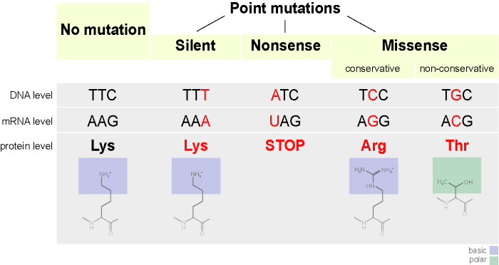
An illustration of three types of point mutations to a codon. (Source: Wikipedia-CC BY-SA 3.0)
Sickle cell anemia is a genetic disease caused by an incorrect codon of one amino acid in a protein such that Valine replaces glutamine as the sixth amino acid in one of the protein changes coded for by DNA. Such a small change has a serious impact on the formation of red blood cells, causing them to have a sickle shape, which reduces their oxygen carrying capacity.
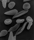
A scanning electron micrograph showing a mixture of red blood cells, some with round normal morphology, some with mild sickling showing elongation and bending. (Source: Wikipedia-CC BY-SA 3.0)
Cancer is a condition characterized by uncontrolled cell division. Cancer can be caused by nitrogen base substitution, or the movement of genetic material from one part of the chromosome to another. There are many cancer causing agents (carcinogens) including X rays, UV radiation & mutagenic chemicals and viruses, that promote the alteration of normal genes into cancer causing genes (oncogenes).
Mitosis is a type of asexual reproduction, cell division results in 2 identical 'daughter' cells being produced from a 'parent' cell. An average human being has about 60 trillion cells and millions of cells are constantly dividing to maintain this number. Some cell types divide faster (such as hair cells), relatively slowly (such as stomach epithelium) and others do not divide at all (nerve cells). Mitosis is necessary for growth, tissue/cell replacement and for tissue repair (after injury).
Mitosis is a continual process, but can be divided into 4 phases.
Prophase
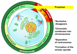
A detailed illustration of the Cell Cycle and Prophase. (Source: Wikipedia-CC BY-SA 3.0 / Public Domain)
Metaphase
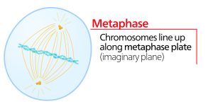
An illustration of Metaphase, and most specifically the formation of the metaphase plate. (Source: Wikipedia-CC BY-SA 3.0)
Anaphase
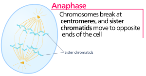
An illustration of Anaphase. (Source: Wikipedia-CC BY-SA 3.0)
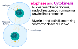
Telophase
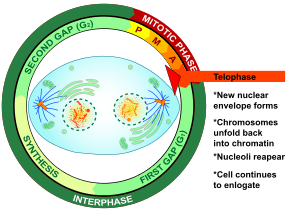
An illustration of Telophase. Nuclear membranes begin to form around two separate sections of what will become two separate cells. (Source: Wikipedia-CC BY-SA 3.0)
Cytokinesis
The Cytoplasm begins to divide by forming a cleavage furrow at the equator and pinches off forming 2 daughter cells, with genetic information identical to each other. These cells will become the new parent cells for the next cell division cycle.
An illustration of Cytokinesis with two cells formed, marking the end of Mitosis. (Source: Wikipedia-CC BY-SA 3.0)
Mitosis - Animals versus Plants
There are 2 main differences between animal and plant cell division:
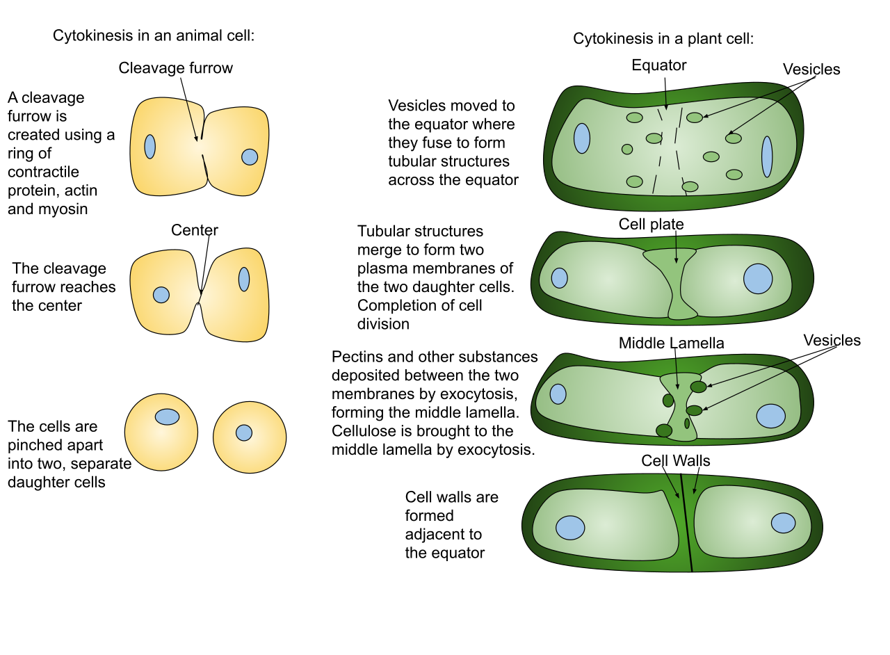
A detailed illustration of the differences between Cytokinesis in animals and in plants. (Source: Wikipedia-CC BY-SA 3.0)
Cloning is the process in which identical offspring are formed from a single cell or tissue. Many plants have the ability to naturally clone themselves; this is called totipotent (i.e. strawberries, aspen trees). Most animal cells are not totipotent (except salamanders) but technology has advanced to allow us to clone (i.e. Dolly the sheep).
Cloning techniques include nuclear transfer from blastula, nuclear transfer with electrical or chemical shock, identical twins.
Nuclear transfer from blastula:The nucleus of a cell from a blastula is removed and transplanted into an enucleated unfertilized egg. Enucleated cells are cells that do not contain a nucleus. The egg begins to divide by mitosis, eventually forming an organism.
Identical twins: Occur when a zygote divides into two and each undergoes successive mitotic cycles to form the multicellular embryos The genetic information between the two embryos is identical
The average cell will divide between 50 and 70 times before cell death. As the cell divides, the telomeres on the end of the chromosome get smaller. If cells divided without these telomeres, they would lose the ends of their chromosomes, and the necessary information they contain. Telomeres are disposable buffers blocking the ends of the chromosomes, are consumed during cell division, and are replenished by an enzyme, telomerase reverse transcriptase. The Hayflick limit is the theoretical limit to the number of times a cell may divide until the telomere becomes so short that division is inhibited and the cell enters senescence.
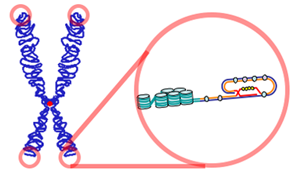
An illustration of the telomeres located at the tips of chromosomes. (Source: Wikipedia-CC BY-SA 3.0)
Karyotypes describe the chromosome count of an organism and what these chromosomes look like under a light microscope. Details provided include their length, the position of the centromeres, banding pattern, any differences between the sex chromosomes, and any other physical characteristics.
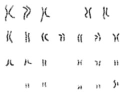
The Karyotype of a male individual showing a smaller Y chromosome. (Source: Wikipedia-CC BY-SA 3.0)
Meiosis is a type of cell division that results in the formation of gametes (sex cells). Gametes may be either sperms or ova (eggs). Gametes contain half the genetic information of other body cells, and are thus described as Haploid. Each sperm or egg produced carries 1 of over 8 million possible combinations of parental chromosomes. A human germ cell with 46 chromosomes will undergo meiosis and produce gametes that have 23 chromosomes. The 46 chromosomes number is referred to as diploid and is written as 2n. The 23 chromosomes number is referred to as haploid and is written as n. Fertilization occurs when 2 gametes (sperm & egg) fuse, forming a diploid zygote (46 chromosomes).
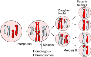
An overview of Meiosis. In general, Meiosis occurs in two stages, Meiosis I and Meiosis II. (Source: Wikipedia-CC BY-SA 3.0)
Meiosis occurs in two main phases namely Meiosis 1 and Meiosis 2. Meiosis 1 comprises of Interphase, Prophase I, Metaphase I, Anaphase I and Telophase I. These are followed by Meiosis 2, which comprises of Prophase II, Metaphase II, Anaphase II and Telophase II.
Interphase
As in mitosis, interphase (cell growth and DNA replication), must occur before cell can replicate. Interphase occurs before prophase I but not before prophase II.
Prophase I
Metaphase I
Anaphase I
Telophase I
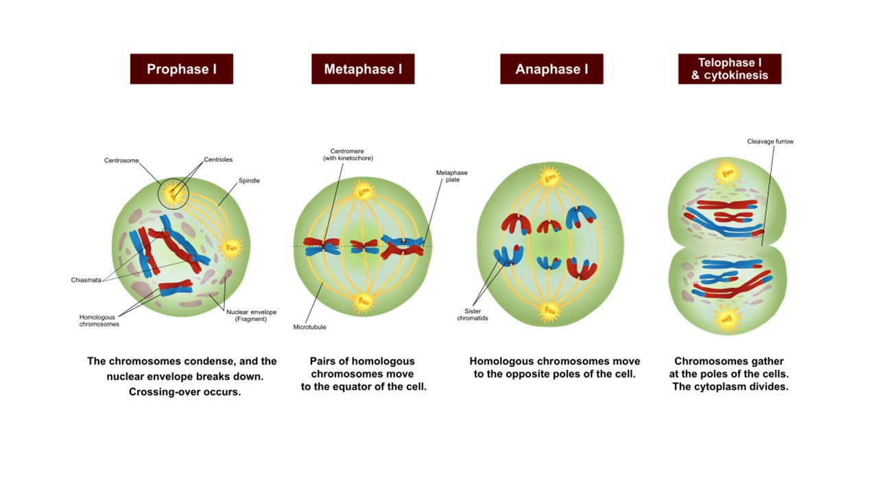
An overview of Meiosis I. (Source: Wikipedia-CC BY-SA 3.0 - Edited by Foundations Learning Hub staff to optimize the screen)
The short phase between Meiosis I and II is referred to as interkinesis. There may or may not be an interphase II depending on species. The next set of cell divisions will separate the chromatids.
Prophase II
Metaphase II
Anaphase II
Telophase II
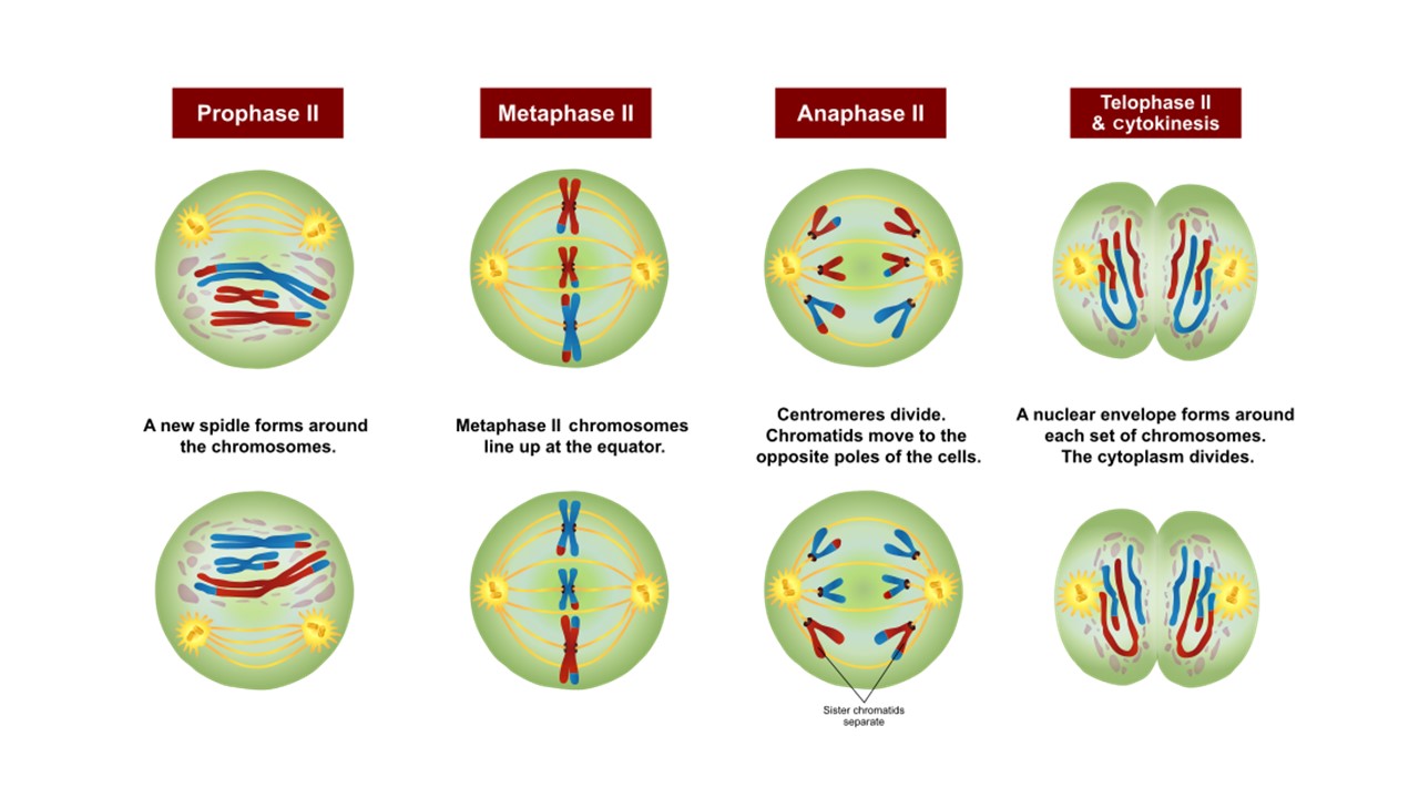
An overview of Meiosis II. (Source: Wikipedia-CC BY-SA 3.0 - Edited by Foundations Learning Hub staff to optimize the screen)
It is important to remember that Meiosis I is referred to as reduction division because the chromosome number is reduced by half. Meiosis II is called equational division and is similar to mitosis as centromeres on sister chromatids separate and chromosome number remains unchanged.
Phenotype: Also called trait, or character. It is the observable/detectable and measurable characteristic of the organism.
Genotype: The genetic composition of an organism at a specific locus. A genotype at a specific allele consists of two letters, one representing the gene from the mother and the other representing the gene from the father. If the two genes are the same, the genotype is described as homozygous, while if the two genes are different, the genotype is heterozygous.
Locus (many=Loci): The exact location of the gene on the chromosome.
Chromosome: A thread-like structure made up of the DNA strand and histone proteins. Chromosomes appear in pairs called homologous chromosomes with one chromosome of each pair coming from either of the parents. Humans have 46 chromosomes, or 23 pairs of chromosomes. At the end of DNA replication, chromosomes appear to have two chromatids, joined at the centromere.
Allele: Alternate forms or varieties of a gene.
A Carrier: An individual who is heterozygous for a trait that is dominant. This individual can pass their recessive alleles to their offspring and cause the phenotype to be observed in the offspring.
Dominant allele: An allele that masks the presence of a recessive allele in the phenotype. So that the phenotype expressed by the homozygous dominant (AA) and the heterozygous genotypes (Aa) will be the same.
Recessive allele: Is the masked allele. Recessive alleles are expressed in the phenotype when the genotype is homozygous recessive (aa).
Pleiotropy: A situation when a single gene is responsible for the expression of multiple traits.
Hybrids: Offspring that are the result of mating between two genetically different parents. Thus, it is the opposite of purebred.
Incomplete penetrance: Is the phenomenon where an allele is expressed only if certain conditions are present in the environment.
Polygenic trait: A trait influenced by multiple genes. Simple Mendelian rules of dominance do not apply to the complex interaction of these genes. As a result, phenotypes may appear as apparent blends or intermediate expressions. Good examples of polygenic traits are human skin and hair color. Many polygenic traits are also influenced by environmental factors.
Heredity is the study of the passing of traits and genes from parents to offspring. Biological traits are controlled by genes. Therefore, a gene is the basic unit of heredity. There is only one copy of the same gene in a chromosome.
Genes are contributed by each parent and the combination of the genes results in the trait that can be observed.
Gregor Mendel, described as the father of heredity, was an Austrian Monk who lived from 1822 to 1884. He conducted several experiments at the Monastery using the garden pea.
Gregor Mendel, 1822-1884. (Source: Public Domain)
The garden pea was an ideal plant to use for Gregor's experiments because:
Gregor Mendel chose to work with 7 characters or traits including:
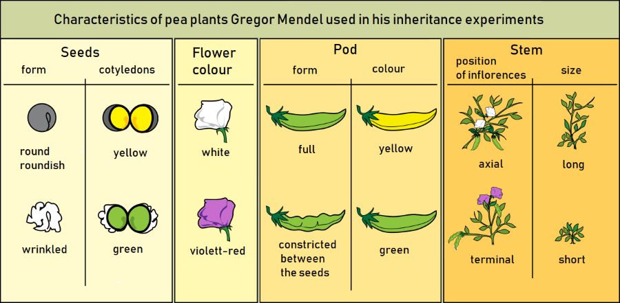
The characteristics used in Mendel's pea experiments. (Source: Wikipedia-CC BY-SA 3.0)
A cross between purebred round (RR) seeds and purebred wrinkled seeds (rr) resulted in all round seeds (expected offspring genotype Rr). He then crossed the F1 (first filial) hybrids (Rr) to produce an F2 (second filial) generation and observed that 75% were round and 25% of the seeds were wrinkled. This resulted in a 3:1 ratio. In this case, the round trait was described as dominant and the wrinkled trait as recessive. In the F2 generation, 50% of the offspring were genotypically heterozygous, but phenotypically round.
First Law (Law of Unit Characters)
Inheritance is governed by genes that exist in the individual and are passed on to offspring. These factors (genes) occur in pairs, one gene comes from the female and one gene comes from the male. The alternate forms of the same genes are known as alleles.
Second Law (Law of Dominance)
One factor, or gene, masks the effect of another. This process is known as the principle of dominance. The dominant expression is seen and the recessive gene is not seen (remains hidden).
Third Law (Law of Segregation & Recombination)
A pair of factors (genes) segregate/separate during the formation of sex cells (meiosis). As a result, each parent can only contribute one member (allele) of a pair of genes to their offspring.
In some texts, the law of segregation, described above, is defined as the First law, and the second law is "The law of Independent Assortment, which describes that genes assort independently and one gene has no influence on another gene during gametogenesis.
A monohybrid cross focuses on one trait. For example, in this case plant height as either Tall or short. Cross 2 contrasting traits e.g. Tall (TT) and Short (tt), all the offspring will be heterozygous Tt, and therefore Tall. A Punnett square can help organize the results of a cross between the sex cells of two individuals. Using the punnett square, we can predict the genotypes & phenotypes of the offspring. Because phenotypes do not always correspond with distinct genotypes, phenotypic ratios in a population might differ from genotypic ratios.
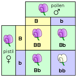
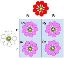
A Punnett square of a monohybrid cross for flower color. The purple color is dominant, and flowers appear purple when they are homozygous dominant BB and heterozygous Bb. Only the homozygous (double) recessive genotype bb results in white flowers. The phenotypic ratio is 3:1, the genotypic ratio is 1:2:1. (Source: Wikipedia-CC BY-SA 3.0)
Dihybrid cross is a cross between two different lines/genes that differ in two observed traits. For genes on separate chromosomes, each allele pair showed independent segregation. If the first filial generation (F1 generation) produces four identical offspring, the second filial generation, which occurs by crossing the members of the first filial generation, shows a phenotypic ratio of 9:3:3:1.
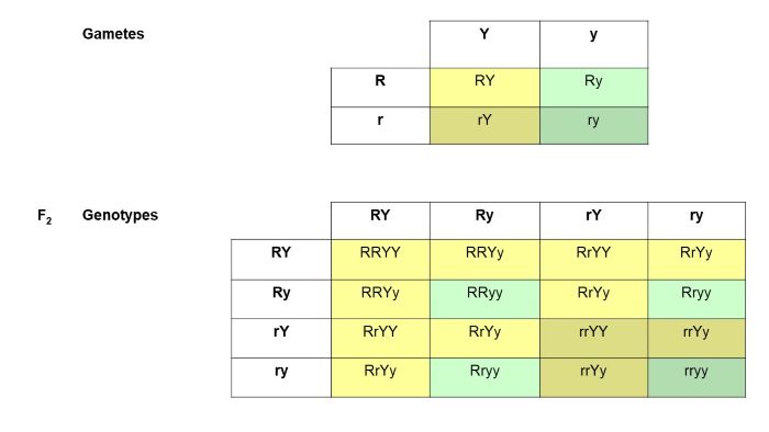
A dihybrid cross focusing on two traits represented by the R or r and Y or y gametes. The gametes table shows all the possible combinations of alleles. Then the second table shows all the possible offspring that can be produced if you had parents with all the possible gametes. This also assumes the upper case represents dominant alleles therefore some individuals will have the same phenotypes even though their genotypes maybe different (such as RRYY and RrYy). (Source: Wikipedia-CC BY-SA 3.0)
A test cross is often performed to determine the genotype of a dominant phenotype. For example, if the white allele is dominant and a farmer selected a white ram and mates with the whole flock, all offspring will be white if the ram is homozygous, but some offspring will be black if the ram is heterozygous. A test cross is always performed by mating the unknown genotype with a homozygous recessive known genotype. If 50% of the offspring are black and the other half white, the unknown genotype must have been heterozygous (Ww). If 100% of the offspring are white, then the unknown genotype must be homozygous dominant (WW).
There are three main types of gene interactions:
There are many traits that are controlled by more than two alleles for example, eye color in Drosophila is controlled by four possible alleles. The following phenotypes and dominance hierarchy is possible: wild type (red) > apricot > honey > white. When dealing with multiple alleles, it is no longer necessary to use upper- & lower-case letters, both letters & numbers are used.
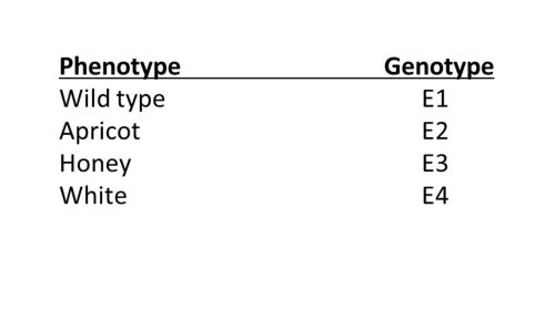
An illustration of eye color multiple alleles in Drosophila.
In some heterozygotes, both alleles of a pair are expressed in the phenotype. These alleles are said to be equally dominant. This lack of dominance is known as incomplete dominance. In this case, a heterozygote of a black and white cross results in grey offspring. Crossing two heterozygous parents would result in a ratio of 1:2:1 where the 2 represents the heterozygous genotype. This should be contrasted with dominant cross, which would have resulted in a 3:1 phenotypic ratio.
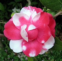
An illustration of Incomplete dominance with pink flowers arising from white and red parents. (Source: Wikipedia-CC BY-SA 3.0)
Another example of incomplete dominance is Sickle cell anemia, caused by a mutation in the Hemoglobin Subunit Beta (HBB) gene. Homozygous normal individuals have optimal oxygen transport and are susceptible to Malaria. Heterozygous individuals (carriers/sickle cell trait) have some difficulty with oxygen transport and some resistance to malaria. Homozygous recessive individuals (sickler / sickle cell disease) have severe difficulty with oxygen transport and are malaria resistant.
A form of incomplete dominance where both alleles are fully expressed in such a way that the effect of each is noticed separately in the phenotype. Example - A red bull crossed with a white cow results in a roan calf (calf has intermingled white & red hair). The ABO blood group system is another example.
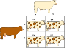
An illustration of Codominance with the offspring expressing the colors of both parents. (Source: Wikipedia-CC BY-SA 3.0)
The ABO Blood Groups Example
The Blood types present in humans are A, B, AB and O. The genotypes that can result in these blood groups are as follows:
Blood group A can result from genotypes AA and AO. Blood group B can result from genotypes BB and BO. Blood group AB results from genotype AB. And Blood group O results from genotype OO. Therefore allele A and B are codominant.
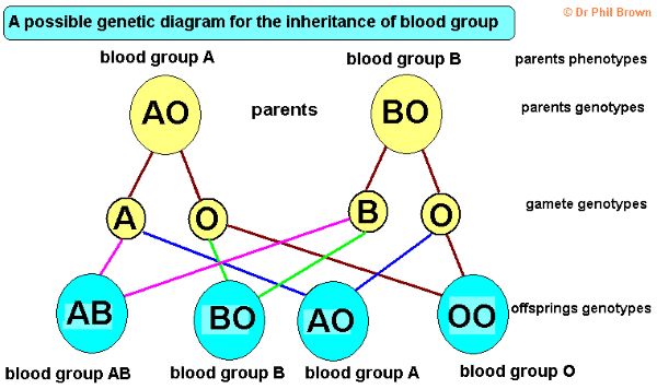
An illustration of Codominance in the ABO Blood group system. (Source: Copyright Dr. Phil Brown)
In each of the 4 distinct blood groups, the Rhesus Protein is either present (+) or absent (-). The inheritance of the Rhesus protein follows a dominant fashion, where the (+) allele is dominant over the (-) allele. Combining the blood type (A, B, AB or O) with the Rhesus factor (+ or -) results in a typical dihybrid cross.
In addition to their role in determining sex, the sex chromosomes, especially X chromosomes, have genes for many characters unrelated to sex. There are very few genes on the Y chromosome that are common on the X chromosome, and because of that, little crossing over may occur between an X and a Y chromosome. Genes located on the sex chromosomes are called sex-linked traits. Female cells show dark spots of chromatin (called Barr Bodies) during interphase, male cells do not. Female cells contain 2 X chromosomes and males contain one X and a Y chromosome, resulting in the genotypes XX and XY for females and males respectively.
Examples of sex linked traits include:
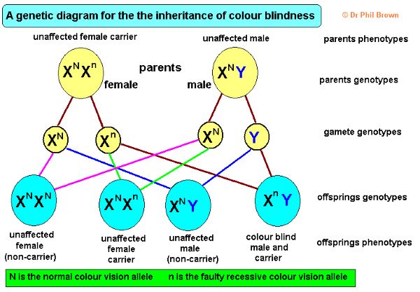
An illustration of the inheritance of sex linked color blindness. (Source: Copyright Dr. Phil Brown)
A chart or register showing a line of ancestors Circles represent females, squares represent males, solid circles & squares represent those who have the trait being studied. Horizontal lines between circles and squares represent mating between male and female. A vertical line joins parents and children. Pedigrees are often used to study sex-linked traits such as color-blindness and hemophilia.
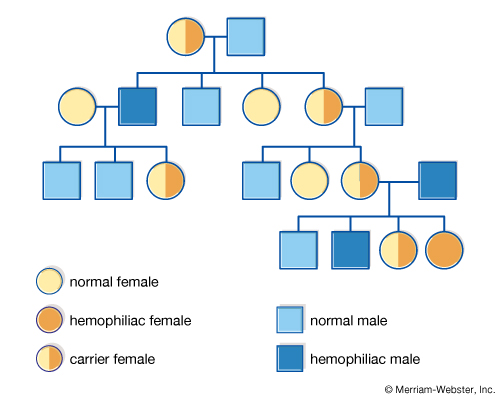
A pedigree diagram with the meaning of the various symbols. (Source: Merriam-Webster, Inc)
To determine if a trait in a pedigree is autosomal or X-linked:
Autosomal Dominant
Autosomal Recessive
X-linked Dominant
X-linked Recessive
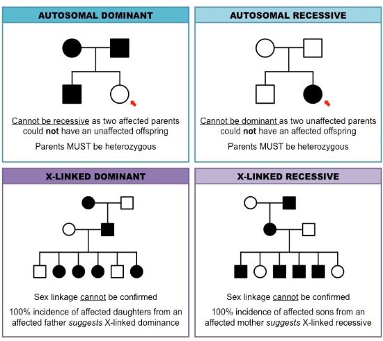
Patterns of inheritance of various types of traits in the pedigree. (Source: https://ib.bioninja.com.au/)
Traits controlled by multiple genes are referred to as Polygenic traits (as compared to Monogenic traits). Genes that interfere with the expression of other traits are called Epistatic genes.
For example - Coat color in dogs: Allele B produces a black coat color in dog, while allele b produces a brown coat color. A second gene, W, prevents the formation of pigment, while w does not prevent color formation. Genotype wwBb would be black, genotype WwBb would appear white because the W allele masks the effect of the B color gene.
Occurs when two different genotypes interact to produce a phenotype that neither is capable of producing by itself.
For example, comb structure in chicken: Allele R produces a rose comb in chickens. Allele P (on a different chromosome) produces a pea comb. Alleles R and P, when present together result in a walnut comb. The absence of rose and pea alleles results in an individual with a single comb.
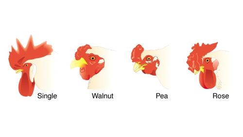
The genetics of chicken combs.
Download a pdf copy of Unit 9 - Cell Division Genetics and Molecular Biology for offline use.
C$0.99
You can access Tensai High School Biology revision questions HERE