Nervous System
Human survival relies on a system of communication between the internal environment and the external environment. Both the nervous system and the endocrine system (hormones) are involved is the communication between the internal and the external environments. This communication is made possible by the electrochemical messages relayed from the brain (nervous system) or by chemical messengers (hormones/blood - endocrine system).
The main function of the nervous system include:
There are two main types of nerve cells found in the nervous system:
Glial cells are neurological cells, which are non-conducting and used for structural and nutritional support.
Neurons are cells that conduct/transmit nerve impulses.
Neurons are the basic unit of the nervous system. Individual neurons connect to one another to form a nerve. Neurons are composed of a cell body and a number of 'branch-like' extensions called dendrites.
Parts of a neuron:
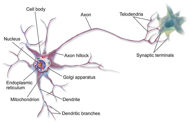
Parts of a neuron. (Source: Wikipedia-CC BY-SA 3.0)
Anaxonic neurons: Found in the central nervous system. As the name suggests, they do not have axons, and their functions are unknown.
Unipolar neurons: This type of neurons have a cell body off to one side. Axons and dendrites are continuous. The neurons are associated with receptors sensing environment i.e. sensory neurons.
Bipolar neurons: This type of axon has a single dendrite and axon with the cell body located in the middle of the axon. They are rare but good examples include the rods and cones found in the eye.
Multipolar neurons: These have several short dendrites and a single long axon with one or more branches. They are most common and they activate glands or muscles, i.e. motor neurons.
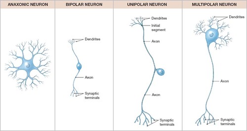
Types of Neurons. (Source: Wikipedia-CC BY-SA 3.0)
An action potential is an electrical and chemical (electrochemical) response generated by neurons as a response to stimuli.
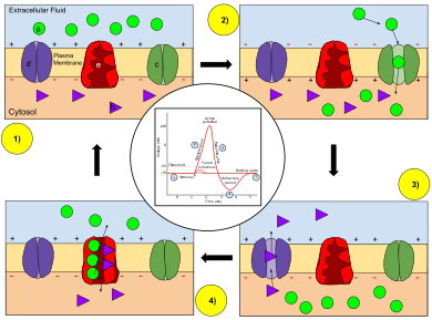
Neurons and other somatic cells have a potential difference (the difference in electric potential between two points) that is established because the cytoplasm is relatively more negatively charged than the extracellular space. This slight negative charge is approximately -70mV and is called the resting potential or membrane potential. The resting potential is caused, and maintained, by the relative concentrations of sodium ions (Na+) and potassium ions (K+). The concentration of these ions is controlled by the Sodium-Potassium (Na+K+) pump, which pumps 2 potassium ions into the cell for every 3 sodium ions that leave the cytoplasm.
An action potential can be broken into three main phases. The cells starts at its resting membrane potential where there is no stimuli, the cell is described as polarized. The three phases of an action potential are Depolarization, Repolarization and Refractory period (due to Hyperpolarization).
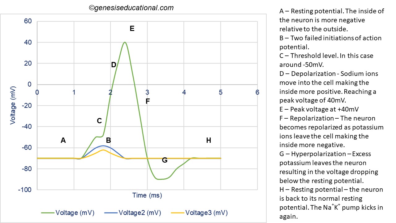
Approximate plot of a typical action potential shows its various phases as the action potential passes a point on a cell membrane. (Source: Wikipedia-CC BY-SA 3.0)
Depolarization: The stimulation of the neuron by electrical discharge (or by certain chemicals) causes sodium ion gates to open and therefore the axon membrane allows sodium ions to pass through the plasma membrane into the cytoplasm. Sodium ions rush into the axon resulting in a positive charge on the inside of the axon relative to the outside. This changes the membrane potential from -70mV to +40mV, and the membrane is said to be depolarized. Once the sodium ions establish an equilibrium, the sodium ion gates close and the membrane becomes impermeable to them again.
Repolarization: At the peak of the action potential, once the sodium ion gates close, the potassium gates open and potassium moves outside the cell. This results in the cytoplasm becoming negatively charged again but this time with the sodium and potassium ions on the wrong sides of the plasma membrane. The (Na+K+) pump will then kick in and exchange the sodium ions and potassium ions to restore the resting potential. This process of re-establishing the resting potential is called Repolarization.
Until the resting potential has been properly restored, a subsequent action potential cannot be conducted along the axon. The amount of time taken to restore the resting potential is called the refractory period. The refractory period is about 1 ms but the stronger the impulse, the longer it takes for the nerve to recover.
In myelinated neurons (wrapped with Schwann cells) the nerve impulse jumps from one Node of Ranvier to the next, greatly speeding up the transmission. This type of transmission is called Saltatory Conduction.
Sodium and Potassium ion movement during an action potential. (Source: Wikipedia-CC BY-SA 3.0)
The nerve impulse travels through the entire length of the axon until it reaches the axon terminal. This terminal is in close contact with the dendrites of the next neuron. The space between the axon of the neuron and the dendrites of the next cell is called a Synapse. The axon terminal contains numerous synaptic vesicles and mitochondria, which supply energy to the cell. The vesicles store chemical transmitters (also called neurotransmitters), which act as messengers that move through the synapse to help carry the impulse from one neuron to another.
The Presynaptic neuron is the neuron that carries the action potential that is transferred across the synapse. The postsynaptic neuron is the neuron that will receive the action potential after it crosses the synapse. The synaptic cleft is the small space between the presynaptic and the postsynaptic neurons. The end of the presynaptic neuron (synaptic end bulb) has synaptic vesicles that contain neurotransmitters. The postsynaptic neuron has neurotransmitter receptors on its cell membrane, which are essential in binding the neruotransmitter.
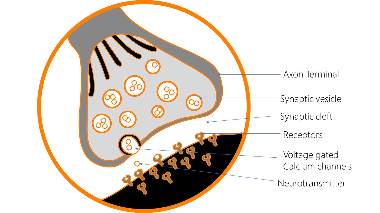
A general illustration of a chemical synapse.
When an action potential arrives at the synaptic end-bulb, it causes voltage gated calcium gated channels to open, calcium passes through these gates and enters the presynaptic neuron. Once inside, calcium binds onto the synaptic vesicles and causes the vesicles to fuse to the presynaptic membrane. The neurotransmitter is released into the synaptic cleft and binds onto the neurotransmitter receptors on the membrane of the postsynaptic neuron. The neurotransmitter causes depolarization of the postsynaptic membrane creating an action potential. The neurotransmitters are now released from the receptors and may either be broken down by an enzyme, such as acetylcholinesterase, or they may move back into the presynaptic neuron and form vesicles again awaiting the next action potential.
Neurotransmission can also occur through electrical synapses as a result of the direct connection between excitable cells in the form of gap junctions, an action potential can be transmitted directly from one cell to the next in either direction. The free flow of ions between cells enables rapid n on-chemical-mediated transmission.
Neurotransmitters can be classified into two major types; excitatory or inhibitory.
Excitatory Neurotransmitters: The main excitatory neurotransmitter is Acetylcholine (ACh). Acetylcholine is released into the synaptic cleft and facilitates the transmission of an action potential by causing the sodium channels of the post-synaptic membrane to open. Once the postsynaptic neuron is depolarized, the enzyme cholinesterase (or acetylcholinesterase) is released to break it down so that the it doesn't continually stimulate the post-synaptic neuron. Some of the ACh is taken back to the presynaptic neuron. Acetylcholine is essential for alertness, learning and memory. Other examples of excitatory neurotransmitters include norepinephrine (Noradrenaline) and epinephrine (Adrenaline).
Inhibitory Neurotransmitters: The main examples are Gamma Aminobutyric Acid (GABA) and Serotonin. Once they are released at the synaptic cleft, they cause hyperpolarization of the postsynaptic membrane, which prevents an action potential from developing. They do this by making the postsynaptic membrane more permeable to potassium ions, which flow outside the cell, resulting in an even lower negative charge. Malfunctions associated with GABA are often common in people suffering from epilepsy, Huntington's disease and some people with sleep disorders.
A neuron will either fire, or not fire. This is called the All-or-None principle. A stimulus must be large enough to start an impulse, i.e. it must reach a threshold level. The threshold is the minimum stimulus needed to produce an action potential. Different neurons have different thresholds, but on average the depolarization has to reach around -55mV. The intensity of a stimulus can be detected by increases in the frequency of the nerve impulses. Increasing the strength of the stimulus does not increase the impulse strength. This explains the nerve impulse in response to touching a warm glass rod versus touching a hot glass rod, with the hot glass rod causing impulses at a higher frequency.
Summation, in Biology, is the additive effect of several electrical impulses. Successive stimuli on one nerve are called temporal summation, while the addition of simultaneous stimuli from several conducting fibers is called spatial summation. Summation can be divergent or convergent. Divergent summation is when one neuron connects to multiple neurons, which might further connect to even more neurons. Convergent summation is when multiple neurons connect to successively less neurons and might end up connecting to just one final neuron. In general, Summation of excitatory postsynaptic potentials increases the probability that the potential will reach the threshold potential and generate an action potential, whereas summation of inhibitory postsynaptic potentials can prevent the cell from achieving an action potential. The concept of threshold potential means there are several stimuli that may not be detected because they did not trigger an action potential. These have been shown in the figure above as 'failed initiations'.
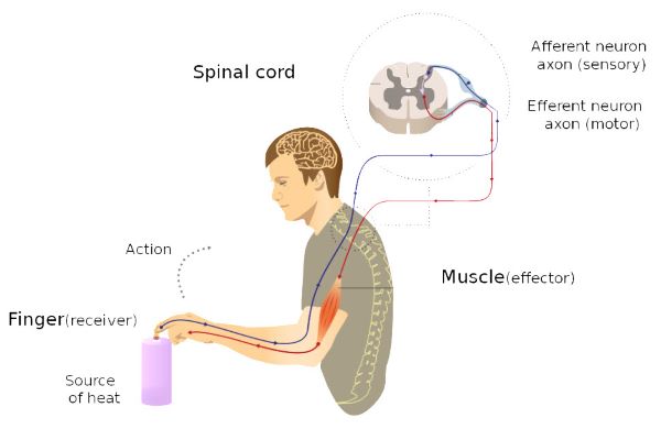
Reflexes are involuntary (autonomic), rather than voluntary (somatic), responses. They are also called reflex actions. They are the simplest forms of sensory stimuli because they do not involve brain stimulation. The pathway of neurons that are involved is called the reflex arc. A good example of a reflex action is the knee jerk reflex.
Reflexes have 7 components, which include:
The 7 Components of a reflex arc. (Source: Wikipedia-CC BY-SA 3.0)
The brain is not involved in completing reflex actions, so it only finds out after the response has already occurred. Most reflex responses are protective in nature, i.e. are designed to enable organisms to move away from danger/harm fast. For example the withdrawal reflex that allows you to withdraw your finger from a hot surface.
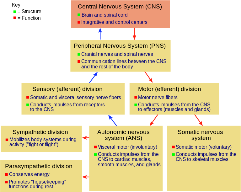
The divisions of the nervous system. (Source: Wikipedia-CC BY-SA 3.0)
The Nervous System can be divided into two major divisions; The Central Nervous System (CNS) and the Peripheral Nervous System (PNS). The Central Nervous System includes the brain and the spinal cord. The Peripheral Nervous System can be further divided into Somatic nerves and Autonomic nerves. Somatic nerves include Sensory and Motor nerves. Autonomic nervous system is categorized into either Sympathetic or Parasympathetic.
The central nervous system is covered by membranes called meninges. These prevent the exposure of brain & spinal cord cells against bone. A fluid called cerebrospinal fluid fills the space between the CNS and the meninges and bathes the spinal cord & brain. The fluid acts as a shock absorber and transports nutrients and wastes to and from the brain. The brain has spaces/grooves/clefts/furrows called fissures, which are also filled with cerebrospinal fluid.
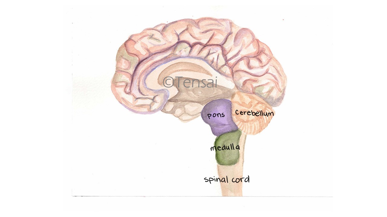
The brain is made up of three main sections including Hindbrain, Midbrain and Forebrain.
The hindbrain consists of the cerebellum, medulla oblongata and the pons.
The cerebellum is located just above medulla. It consists of both grey and white matter. It is furrowed in appearance and is responsible for limb movement, balance and coordination. The cerebellum also organizes and integrates impulses.
The medulla oblongata lies just above the spinal cord, looking like a swollen extension of the spinal cord. In general, it is responsible for the autonomic nervous system (ANS) activities like stimulation of diaphragm, breathing muscles, heartbeat and artery diameter. It is also the pathway for impulses moving from higher parts of brain to motor nerves and muscles.
The pons is the bridge between the cerebellum and medulla oblongata and transmits impulses between the two sides of the cerebellum.
Parts of the hindbrain.
The midbrain is the relay center for some eye and ear reflexes. It contains a network of neurons known as the Reticular Formation that activates the forebrain. It allows you to concentrate on specific stimuli and ignore the rest.
The forebrain consists of the thalamus, hypothalamus and cerebrum.
The Thalamus
The thalamus is a sensory relay center located between the midbrain and cerebrum. It directs incoming sensory signals to the cerebrum. The thalamus has a significant role in sleep, wakefulness, consciousness, sensation of temperature and degree of pain.
The Hypothalamus
The hypothalamus connects to the pituitary gland and controls its functions. The major center for the integration of the nervous & endocrine systems. The hypothalamus controls blood pressure, urine production, water balance (produces ADH), hunger, body temperature, internal organs, autonomic nervous system. The hypothalamus also affects behavioral and emotional responses including rage and sex drive. This pituitary gland on the other hand produces and stores hormones.
The Cerebrum
The Cerebrum is the uppermost and outermost and largest portion of the human brain accounting for about 80% of the brain mass. The cerebral cortex surrounds the cerebrum and is composed of grey matter, the inside of the cerebrum is composed of white matter The cerebrum is also highly folded, which increases the surface area.
The cerebrum is divided into two cerebral hemispheres, the left and right hemispheres, by the Longitudinal fissure. The right side of the brain controls the left side of the body and the left side of the brain controls the right side of the body. The two hemispheres are connected by a bundle of nerve fibers called Corpus Callosum.
A series of fissures divide the brain into four lobes namely Frontal lobe located on the front of the head; Temporal lobe located at the temples (above the ears); Parietal lobe located at the top and back of the cerebrum; and the Occipital lobe located in the lower back of the cerebrum.
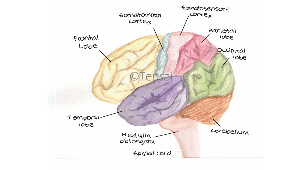
Brain lobes as part of the cerebrum.
The peripheral nervous system comprises of the somatic nerves (sensory and motor nerves) and the Autonomic nervous system. Somatic Nerves are for voluntary control of skeletal muscle, bones and skin. They can be classified in to Motor nerves, which carry messages from the CNS to effectors such as glands or muscles, and, Sensory nerves , which carry messages to the CNS.
The Autonomic Nervous System (ANS) controls involuntary body actions. It can be subdivided in to Sympathetic Nervous System (which prepares the body of emergencies) and Parasympathetic Nervous System (which returns the body to normal after an emergency response).
Sympathetic Nervous System
Causes the adrenal medulla to secrete adrenalin, which prepares body for fight or flight, Adrenaline has the following effects.
The Parasympathetic Nervous System (PNS)
The PNS is generally inhibitory and tries to restore homeostasis after an emergency response. These effects include:
The nervous system receives input from the five senses: touch, taste, smell, sight and sound. Sight, taste, smell and sound are controlled by the autonomic nervous system and are therefore called Special senses. Touch senses, which include pain, temperature and pressure are controlled by the somatic system and are therefore called voluntary senses.
The skin has receptors for pressure (mechanoreceptors), pain (nociceptors), and temperature (Thermoreceptors - hot or cold). These receptors are not evenly spread throughout the skin and they differ in sensitivity. For example, the mouth, tongue, and fingertips are more sensitive to pressure, while the back and chest are least sensitive. This is also similar for temperature receptors. The mouth is able to withstand higher temperatures than other parts of the body. Which explains why hot water feels hotter in the throat than in the mouth. In general, the body has more cold receptors than heat receptors.
The Sensory or Cortical Homunculus is a pictorial representation or a map of brain areas dedicated to sensory processing for different anatomical divisions of the body. The size indicates the sensitivity of the body parts to stimuli.

An illustration of the sensory Homunculus.
Taste is detected by Chemoreceptors. Small bumps on the tongue called papillae and contain taste buds. There are four types of specialized taste receptors: salty, sweet, bitter, sour, and umami. Taste buds are actually buried in the tissue; a small pore lets solutions into stimulate the receptors in the taste bud. Saliva is needed in order to taste because it dissolves molecules.
Taste receptors lead to a variety of different parts of the brain including olfactory bulb, cerebellum, and temporal lobe. Sweet, umami, and bitter tastes are triggered by the binding of molecules to G protein-coupled receptors. Saltiness and sourness are perceived when alkali metal or hydrogen ions make contact with taste buds. Saltiness - alkali while Sourness - hydrogen ions.
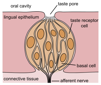
The structure of a taste bud. (Source: Wikipedia-CC BY-SA 3.0)
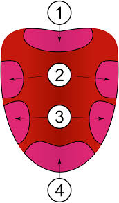
An illustration of the tongue and the regions where specific tastes are detected. 1: Bitter zone, 2: Sour zone, 3: Salty zone and 4: Sweet zone. (Source: Wikipedia-CC BY-SA 3.0)
Olfactory cells are located in the upper part of the nasal cavity and they respond to smell. Olfactory cells are columnar cells with cilia, which act as sensors. The cilia are attached to nerves which lead to the brain.
The eye is a photoreceptor i.e. it detects light of different intensities and wavelengths.
Parts of the eye:
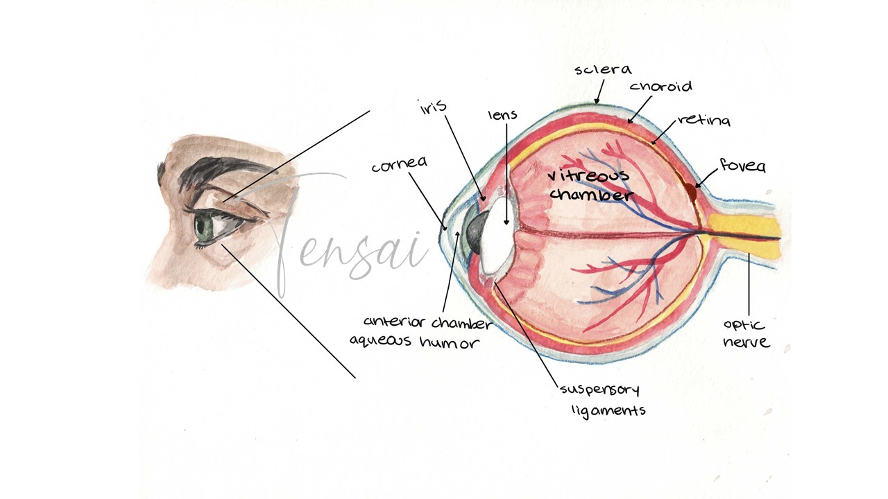
Anatomical structure of the eye.
Rods a very sensitive to light and can be stimulated by even very dim light, making them reponsible for night vision. They rely on a pigment called Rhodopsin that is formed using Vitamin A. When stimulated by light, Rhodopsin breaks down into two small molecules named Retinal and Opsin. This chemical reaction results in a nerve impulse in the sensory neurons attached to the rods. Many rod cells are connected to one sensory neuron so the vision produced by stimulation of rod cells is not very clear. This explains why humans cannot see clearly at night. In addition, Rods are more concentrated in the peripheral regions, away from the macula (the center of the retina) making them (rods) responsible for peripheral vision.
Cones are about 300 times less sensitive to light than rods so they only function under bright light. There are three different types of cones sensitive to one of three colors of light i.e. Blue, Green or Red. Fewer cones are attached to the same neuron, this results in higher resolution in the vision. Cones are also more concentrated directly behind the lens in the fovea centralis.
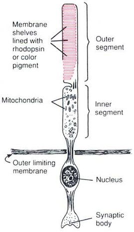
The structure of rods and cones showing slight differences between the two photoreceptors. (Source: Wikipedia-CC BY-SA 3.0)
The human eye can detect light in the 400-700nm range, which really is only a small portion of the electromagnetic spectrum. What makes a color a color is because that wavelength is what is being reflected and every other color is being absorbed. White light is detected when all colors are being reflected, while black color is detected when all colors are absorbed.
Light enters the pupil and passes through the lens onto the retina at the back of the eye. The ciliary muscles stretch or relax to accommodate and focus the lens on the object. The rods and cones in the retina detect the image. Rods are more common in a circular zone, near the edge of the eye while cones occur in the center (fovea centralis) of the retina. Light reaching the rods causes the pigment rhodopsin to break down into retinal and opsin, which in turn causes a membrane potential in the neurons. Cones contain a similar pigment, but are less sensitive to light. This action potential transfers to synapsed neurons that connect to the optic nerve, which in turn connects to the occipital lobe in the brain. It is here in the occipital lobe where the image is translated into information.
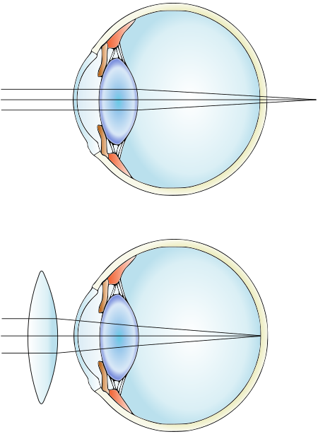
To focus on a near object, the muscles contract and shorten, the lens then becomes more spherical in shape which bends light more and allows the image of the object to be focused on the retina correctly.
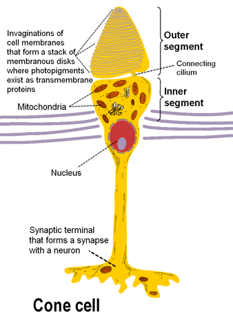
An illustration of astigmatism where the image is distorted. (Source: Wikipedia-CC BY-SA 3.0)
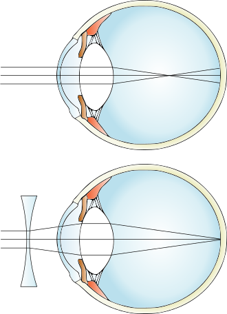
An illustration of short sightedness and correction using concave lenses. (Source: Wikipedia-CC BY-SA 3.0)
An illustration of far sightedness and correction using convex lenses. (Source: Wikipedia-CC BY-SA 3.0)
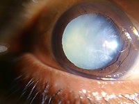
A cataract in a 60 year old individual. (Source: Wikipedia-CC BY-SA 3.0)
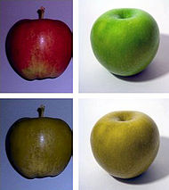
An illustration of color blindness. A normal person sees the two apples as they look on the top row, a color blind person sees them as they look on the lower row. (Source: Wikipedia-CC BY-SA 3.0)


An image of a child suffering from conjunctivitis and an image of an adult with strabismus. (Source: Wikipedia-CC BY-SA 3.0)
Distance Determination can occur in two ways; the brain relates size of objects to the size of known objects to determine size of an object. In addition, the brain can use stereoscopic vision, which is the vision created by the two eyes and the overlapping of vision to can be used to determine distance of an object.
The ear has two major functions, Hearing and Balance. It is made up of three main parts: The Outer Ear, The Middle Ear and The Inner Ear.
The Outer Ear
The main function of the outer ear is to gather sound waves and funnel them to the auditory canal. The outer ear is made up of skin and cartilage. The ear flap is also called the Pinna. The Auditory (ear) canal leads into the head and is lined with wax secreting cells. The ear drum (the tympanic membrane) is the end of the auditory canal and marks the end of the outer ear, and the beginning of the middle ear.
The Middle Ear is home to the three smallest bones in the human body. The Malleus (Hammer), Incus (Anvil) and Stapes (Stirrup). Together, these three bones make up the ear ossicles. The bones are attached to each other by ligaments that allow them to move back and forth and amplify the sound. The last ossicle, the stirrup, is attached to the Oval Window. The Eustachian tube is located just after the eardrum, in the middle ear. It connects to the throat and allows for pressure equalization on both sides of the tympanic membrane.
The Inner Ear is responsible for both hearing (by converting vibrations from the ossicles into nerve impulses) and balance. The Cochlea is the organ that is responsible for hearing (this is the place where neurons attach). The Cochlea is coiled, like a snail shell. It is divided into scala vestibule and scala tympani by the cochlear duct. The cochlear duct contains the Organ of Corti where sound receptors (four rows of hair cells) are found. A fluid (called endolymph) surrounds the Organ of Corti. The utricle, saccule, and semicircular canals are responsible for balance and body position.
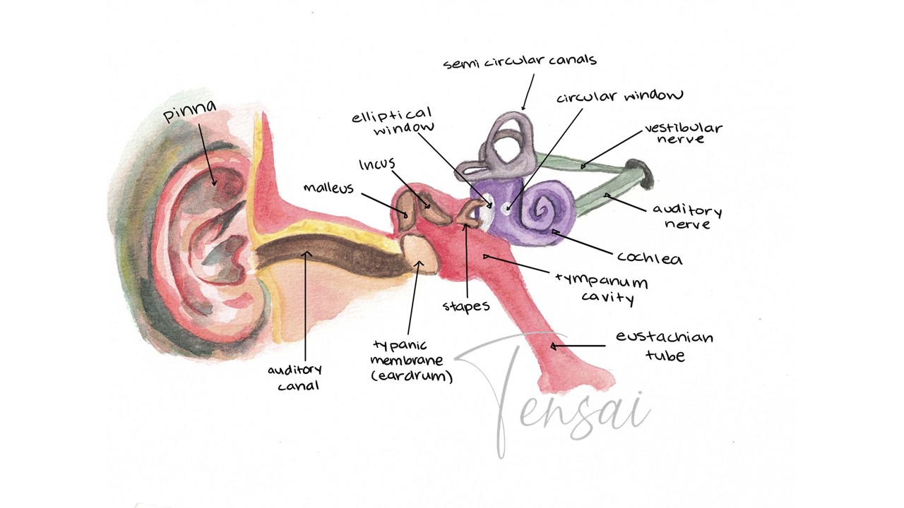
The anatomy of the ear. The brown section represents the outer ear, the red section represents the middle ear, and the purple section represents the inner ear.
Sound waves are funneled by the pinna and travel down the auditory canal causing the Tympanic membrane to vibrate. The ossicles (malleus, incus, & stapes) also vibrate and because the stapes is connected to the oval window, when it moves, it causes this window to bulge. The oval window bulging causes the fluid in the outer two parts of the cochlea to move. This puts pressure on the cochlear duct. The pressure waves cause the basilar membrane to move, which causes the hairs in the organ of Corti to bend against the tectorial membrane. As the hairs bend, the small nerves that are connected to the base of the hairs are stimulated. These nerve impulses are carried to the brain along the auditory nerve to the temporal lobe of the cerebral cortex where the impulses are interpreted into sound.
Sound Intensity and Pitch: Sound intensity depends on the number of neurons and the frequency of firing. The sound Pitch is detectable because each hair cell responds to only one frequency.
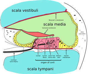
A cross section of the cochlea showing the organ of corti. (Source: Wikipedia-CC BY-SA 3.0)
The utricle, saccule, and semicircular canals are filled with fluid and contain sensory hairs. The utricle & saccule contain small calcium carbonate particles called otoliths (ear stones). Movement of the head, along the horizontal or vertical planes, causes the otoliths to move in response to gravity. These particles cause sensory hairs to bend, triggering impulses, which alert the brain as to the position of the head. One canal detects bending forward and backward, another detects bending left or right, and a third detects a turning motion. These signals reach the brain, and causes stimulation of the appropriate muscles to maintain balance.
There are small muscles in the ears help protect hearing and keep the ear functioning normally. When loud noises are around, the muscles attached to the hammer (malleus) contract and restrict intense movements. A second muscle contracts pulling the stirrup (stapes) away from the oval window limiting inner ear damage. These movements are not effective with sudden loud noises because the response takes time to process. Sudden noise can cause huge damage to the inner ear, specifically the hairs in the Organ of Corti.
Download a pdf copy of Unit 7 - Endocrine and Nervous System for offline use.
C$1.29
You can access Tensai High School Biology revision questions HERE