The Cell
Although we will not go into the details in this text, students are expected to know the contributions from scientists such as:
Magnification: Describes how many times an object is larger than normal. It is expressed using the X symbol, so that if an image is 10 times larger, the magnification is 10X.
Contrast: The extent of light or dark between two structures.
Resolution: The ability to distinguish between two structures that are closely positioned. A high resolution means you are able to clearly tell two closely located structures apart.
Staining: Stains can be used to develop colors to specific parts of the cell to be able to easily identify them. So staining can be used to increase both contrast and resolution.
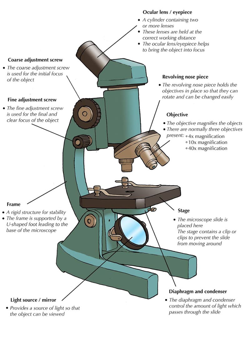
Light microscopes are either Simple (uses a single lens similar to a magnify glass), or Compound uses multiple lenses.
Advantages
Usually inexpensive and provide adequate magnification for large living organisms and structures.
Disadvantages
The maximum magnification is low.
It is used to view specimens that are too think to be viewed by a light microscope. Computers combine several 2D laser scanned images into a 3D image.
Advantages
Higher magnification compared to light microscopes. The 3D images are better representations of true image than 2D rendering.
Disadvantages
There are limited laser wavelengths that can be used.
EMs use a beam of electrons to illuminate specimens. They can achieve a much higher magnification of X200, 000. There are two types of Electron microscopes: Transmission and Scanning.
The beam of electrons produces a 2D image. Specimens need to be processed in a special manner; they are sliced ultrathin and placed in a vacuum.
SEM produces 3D images of the surface of the specimen.
Disadvantages of Electron Microscopes
Electron microscopes can only be used to observe non-living organisms. In addition, the specimens need to undergo extensive processing and preservation.
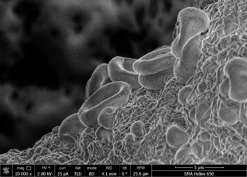
A scanning electron micrograph of a blood clot with red blood cells visible. (Source: Wikipedia-CC BY-SA 3.0)
A fine metal probe is brought near the specimen. Electrons flow between the tip of the probe and the specimen's surface. A computer is used to generate a detailed 3D image at an atomic level.
Magnification
Magnification = Ocular power X Objective power.
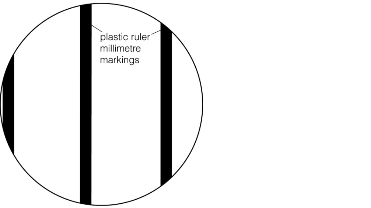
Diameter of the field of view Use a ruler to measure the distance across its center. In the figure below, the diameter of the field of view is 2.5 mm.
Calculating Field Diameter: (Field of View [1]) (Magnification [1]) = (Field of View [2]) (Magnification [2])
All living things are composed of one or more cells. The cell is the smallest basic/functional unit of life.
All living things share specific characteristics, including:
Cells can be classified as either Prokaryotic or Eukaryotic Cells.
Prokaryotic cells are the oldest form of life known. They generally do not have a nucleus. Where the suffix 'karyon' refers to 'nucleus'. Prokaryotic cells include Bacteria, Blue Green Algae, etc.
The genetic material in prokaryotes is sometimes arranged in a circular shape, called a plasmid. Plasmids are very popular for their use in gene cloning.
Prokaryotic cells also do not have organized organelles, though their ribosomes are well organized for protein synthesis. Prokaryotes tend to be smaller than Eukaryotes.
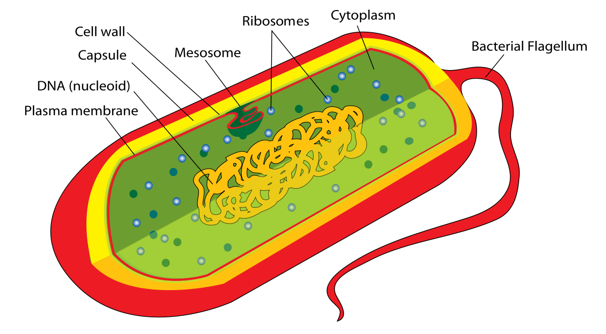
The structure of a Prokaryotic cell. (Source: Wikipedia-CC BY-SA 3.0)
Eukaryotic cells are larger than prokaryotic cells. They have an organized and membrane bound nucleus and organelles.
Eukaryotic cells can either be plant or animal cells. Plant and animal cells differ in:
Purchase a pdf copy of Unit 1 (The Cell) for offline use.
C$0.99
The Nucleus
The nucleus is the brain of the cell. It is a large dark round spot inside Plant & Animal cells. It houses the nuclear DNA and controls the daily activities of the cell. Because it holds the genetic material in the DNA, it can be termed as the library of the cell. Inside the nucleus is a jelly-like fluid medium called the nucleoplasm, that provides support to the contents in the nucleus.
Nucleolus
Inside the nucleus is the Nucleolus. This is a dark spot inside the nucleus and it produces RNA and ribosomes, therefore it is involved in protein synthesis.
Chromosomes
In Eukaryotes, chromosomes are linear and are composed of DNA and proteins. The term 'Chromatin' refers to the mass of stringy, entangled chromosomes observed during interphase (one of the stages in cell divisions).
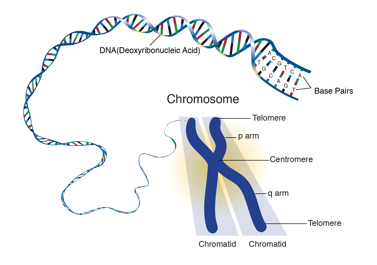
The structure of a chromosome. (Source: nih.gov)
Cytoplasm
Inside the cell, but outside the nucleus is the cytoplasm, which is a gel-like substance that dissolves nutrients to be used for various physiological functions. The cytoplasm also suspends the organelles preventing them from crushing into each other.
Mitochondria
The mitochondria is the powerhouse of the cell. It produces ATP (Adenosine Triphosphate); which is the most common form of energy in the cell. The process involved in energy metabolism is referred to as Cellular Respiration. It is shaped like a sausage on the outside, but contains a double membrane on the inside (outer and inner membrane). The inner membrane is folded/convoluted to increase the surface area for more efficient functioning. The folds are called Cristae.
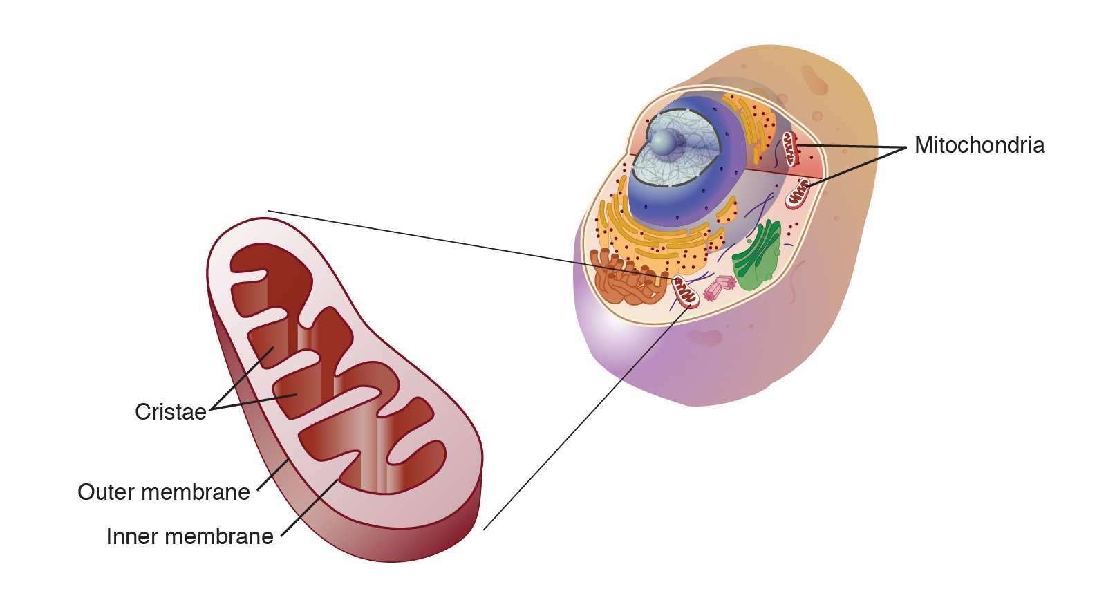
The structure of a mitochondria. (Source: genome.gov)
Vacuole
The vacuole appears hollow when viewed under a microscope. The organelle is more prominent in plant cells and is located somewhat at the center of the cell. Vacuoles function as storage organelles storing water, nutrients and waste.
Ribosomes
Ribosomes are shaped like a snowman, because they contain two subunits. They function in protein synthesis.
Endoplasmic Reticulum (ER)
There are two types of Endoplasmic Reticulum: - Rough and Smooth. The Rough ER have ribosomes attached on the surface and the smooth ER do not have ribosomes attached.
In either case, ER are long tubes and canals in the cell. They have several functions mostly related to transport of molecules.
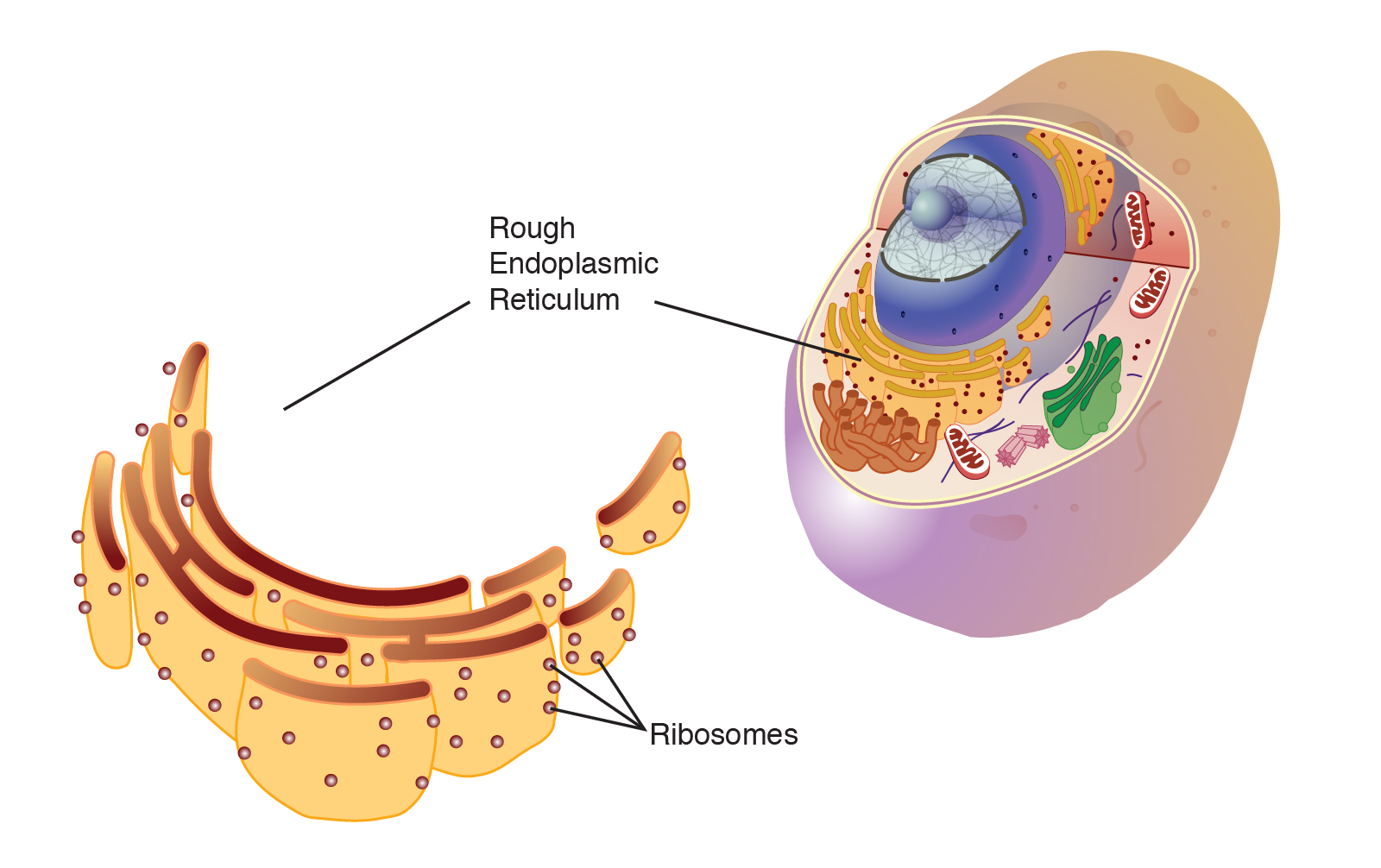
The structure of a rough endoplasmic reticulum. (Source: genome.gov)
Golgi apparatus
Also called the Golgi complex. It looks like a stack of pancakes and is necessary for intercellular transport and for packaging material for transport. It can be likened to a shipping department, receives, sorts and packages various physiological substances for to be used by the cell or sent to other cells.
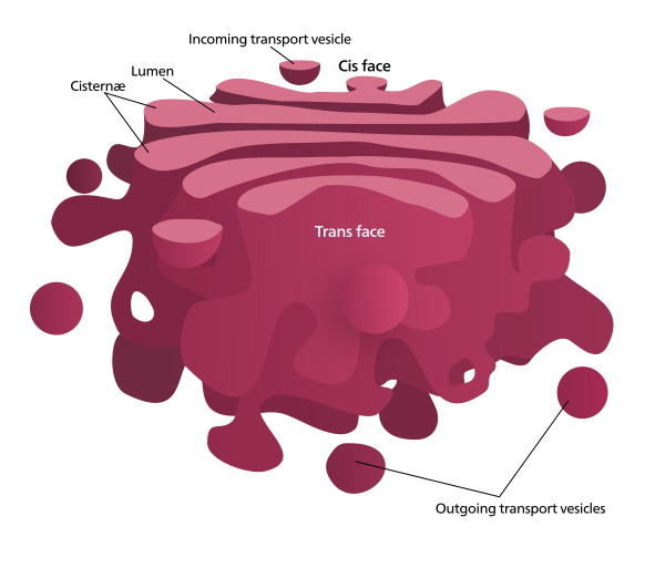
The structure of the Golgi Apparatus. (Source: Wikipedia - under creative commons license.)
Lysosome
Lysosomes are round/spherical bodies that contain lytic enzymes. 'Lysis' means break down. The enzymes are usually proteolytic and require to be contained in lysosomes to prevent the enzymes from breaking down other cellular components.
Centrioles
Centrioles are small rod shaped structures formed by microtubules. They are necessary for cell division.
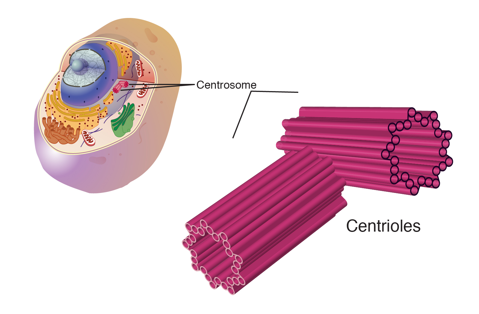
The structure of centrioles. (Source: genome.gov)
Chloroplasts
Chloroplasts are only found in plant cells. They contain chlorophyll which s responsible for capturing light energy for photosynthesis.
Cell Wall
A cell wall is found in plant cells only. It is the rigid thick outer wall surrounding the cell. It gives the cell its regular shape and provides support to the plant. It's made up of cellulose which is also a structural molecule.
Cell Membrane
Also called Plasa membrane. It's present in both plants and animal cells. In plants, the cell wall is located on the inside surface of the cell wall. The cell membrane is made up of a phospholipid bilayer with other large molecules scattered. It retains cell contents inside the cell and allows movement of substances inside and outside the cell.
For cells to perform their physiological functions, substances have to move in and out of the cell at a rate that can meet these physiological requirements. The surface area of the cell is an important factor in influencing the movement of substances in and out of the cell. However, the volume of the cell influences the amount of physiological processes that can occur inside the cell. For instance, larger cells require several more physiological functions to meet the maintenance needs for energy. When the cell grows so big that the cell membrane cannot provide the cell with the materials needed for life processes, the cell divides. The surface area to volume ratio is an important characteristic of organisms. Smaller organisms have a higher SA:V while larger organisms have a lower S.A: V ratio.
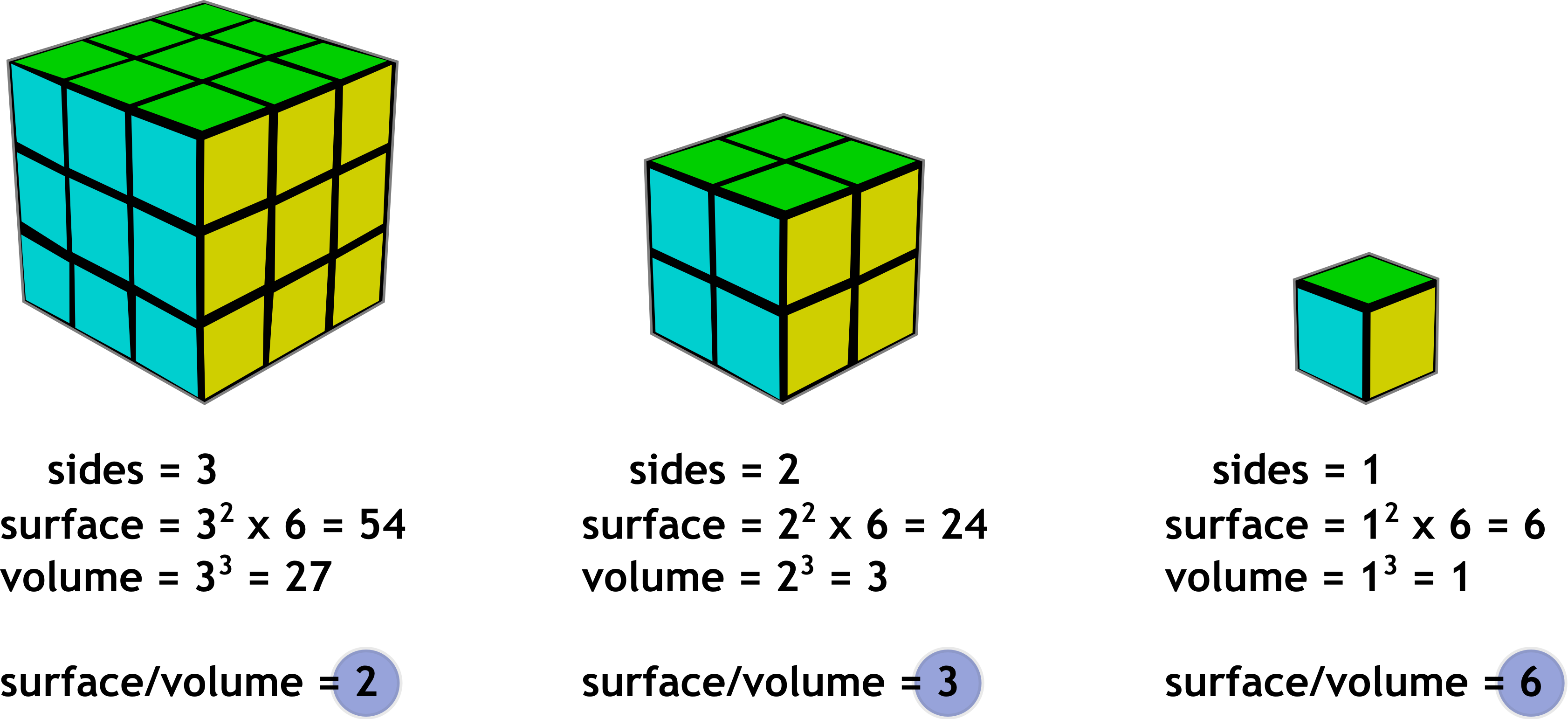
The surface area to volume ratio is an important factor in determining how large a single cell can be, and to predict the metabolic rate of an organism. (Source: Wikipedia-CC BY-SA 3.0)
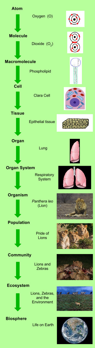
The single cell of a unicellular organism carries out all the functions necessary to keep the organism alive. A group of similar cells that perform the same function make up a Tissue. Animals are mostly composed of 4 types of tissue:
An Organ is a group of two or more types of tissue that work together to carry out a specific function. The skin is the largest organ with several layers made up of different tissues. The heart is also an organ that is made up of muscle tissue, connective tissue and nerve tissue. The brain, lungs and eyes are more examples of organs.
Plants also have organs namely the roots, stem and leaves. These support photosynthesis, absorption of water absorption and transport.
A group of organs working together is called an organ system. The circulatory system in humans combines the heart, blood vessels and blood to deliver oxygen and nutrients to various parts of the body and eliminate waste material. The repiratory system obtains oxygen from the environment and carries it to the blood where it enters the circultory system. Carbon dioxide from the blood enters the respiratory system and is released as a waste product.
A group individuals of the same species living together in a geographical area is a population, such as a pride of lions. Two or more populations interacting with each other form a community. Communities interacting not only with each other but also with the physical environment encompass an ecosystem, such as the Savanna ecosystem. All of the ecosystems make up the biosphere, the area of life on Earth.
The plasma membrane is composed of phospholipids and proteins. The phospholipids are organized in two layers thus referred to as a phospholipid bilayer. As the name suggests, a phospholipid has a phosphate head that is hydrophilic (water loving - can interact with water) and a lipid tail that is hydrophobic (water hating - does not interact with water). Due to this property, the phosphate heads are oriented towards the side of the plasma membrane where water molecules are located.
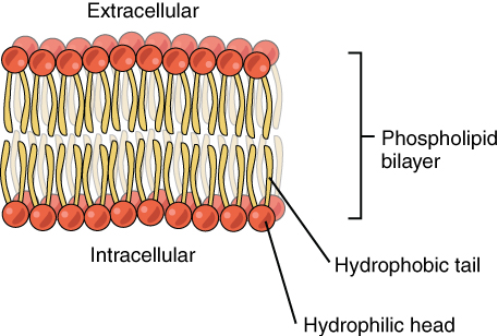
Chemical structure of a phospholipid and the phospholipid bilayer. (Source: Wikipedia-CC BY-SA 3.0)
Retains cell contents
Acts as a barrier, selectively allowing (and blocking) movement of certain material into and out of the cell.
Various proteins are embedded within the two layers of the phospholipids. These are known as membrane proteins. Proteins serve various functions including channels, carriers, signaling, receptor, or enzymes.
Transport across the cell membrane is either Active (requires energy) or Passive (does not require energy to occur).
Passive transport include Diffusion and Osmosis.
Simple Diffusion
The random movement of molecules from an area of higher concentration to an area of lower concentration until the concentration is uniform throughout the space, that's why when you add a drop of red dye into a clear glass of water it will spread throughout the water until it establishes an equilibrium.
Diffusion across a membrane
Membranes can be classified as:
Impermeable: does not let anything pass through the membrane
Permeable: allows all materials to pass through the membrane
Semi-permeable: allows some particles to pass through the membrane while excluding other particles
Diffusion can occur across a semi-permeable membrane, from the side with higher solute concentration (lower water concentration) to the side with lower solute concentration (higher water concentration), until the concentration is equal.
Dynamic Equilibrium
Dynamic Equilibrium describes a scenario where the diffusing particles are still moving, (diffusing) but the movement no longer results in a net change in concentration at one location, more than another location. When there is a membrane, dynamic equilibrium is a state where the diffusing molecules move across the membrane at almost the same rate in either direction.
Concentration Gradient: The difference in concentration between two locations. Molecules of substances move from high concentration to low concentration, so the larger the concentration gradient the faster the diffusion rate.
Temperature: Higher temperature results in faster diffusion rate.
Particle size: The larger the particle the slower the movement.
Facilitated diffusion uses transport proteins to facilitate the diffusion of particles across the plasma membrane. There are 2 types of transport proteins and they are recognized based on their shape, size, and electrical charge: Carrier Proteins are those protein that change shape to allow certain molecules to cross the membrane. Channel Proteins are proteins that form tunnel-like pores in the cell membrane, allowing electrically charged ions in and out of the cell.
Osmosis is the movement of water through a selectively permeable (semi-permeable) membrane from an area of higher water concentration to an area of lesser water concentration.
A solute are molecules that are dissolved in a solvent to form a solution. A solvent is substance that dissolves the solute. In most biological reactions, the solvent will be predominantly water. A solution is the result of dissolving a solute in a solvent.
The environment inside a cell can be described as Intracellular while the environment outside the cell is termed as Extracellular. Usually these two environments differ in their chemical composition.
Based on the relative concentrations (differences) between the intracellular and the extracellular space, three types of solutions can be described.
Hypertonic solution is when there is greater solute concentration outside of cell than inside. In this environment, water will move outside the cell to the extracellular space resulting in the cells shrinking.
Hypotonic solution is when there is lower solute concentration outside the cell, and higher solute concentration inside the cell. Water will move into the cell, the cell will expand, possibly to the point of lysis.
Isotonic solution is when the concentration of molecules is the same inside and outside of the cell. Although water moves in and out of the cell, there is no net change in concentration between the intra- and extra-cellular spaces.
In plant cells, when cells lose water, they shrink and the plant appears wilted. However, in cases where water is entering the cells, the cell walls in plant cells allow them to resist the pressure so they do not burst. This pressure created by water moving into the cells is called Turgor pressure.
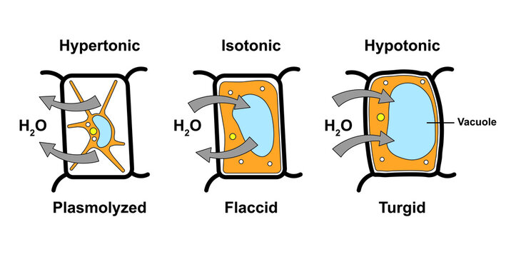
Figure above - The behavior of plant cells under solutions of different tonicity. (Source: Wikipedia-CC BY-SA 3.0)
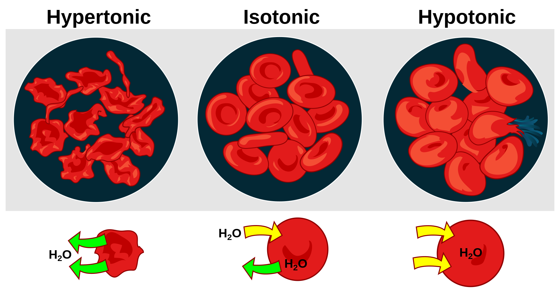
Figure above - The behavior of animal cells under solutions of different tonicity. (Source: Wikipedia-CC BY-SA 3.0)
Active transport can be divided into two main types, active transport protein, or membrane mediated transport. Membrane mediated transport occurs through either Exocytosis (movement outside the cell) or Endocytosis (movement into the cell).
Characteristics of Active Transport
A good example of protein mediated active transport is the sodium potassium ATPase pump.
Cells engulf large particles by extending their cytoplasm around the particle. The engulfed particle is enclosed in a pouch or vesicle, inside the cell.
There are three types of endocytosis
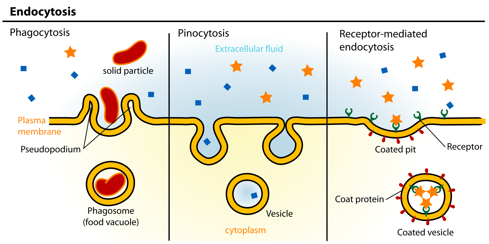
Representation of the different types of endocytosis. (Source: Wikipedia-CC BY-SA 3.0)
Large molecules within the cell are transported to the external environment. A good example is the release of neurotransmitters at a synapse. Vesicles are usually formed at the Golgi apparatus and then move towards the plasma membrane. The vesicle fuses with the cell membrane and vesicle contents are released.
Reverse Osmosis is a process used for water purification where water is filtered by passing it through a membrane with fine pores.
In patients with kidney failure, dialysis is a procedure used to 'clean' their blood. Blood is pumped through tubing made from a synthetic, semi-permeable membrane (dialysis tubing) that is immersed in a salt solution with a concentration similar to blood, but which does not contain wastes. The pores in the tubing allow small dissolved waste molecules to diffuse out of the blood while retaining large proteins and blood cells.
Uses a semi-permeable skin patch, which allows medications to diffuse out of the patch and in to the body at a slow, constant rate.
Plants are multicellular organisms with the ability to create their own food through photosynthesis.
Plants have a variety of specialized structures for different functions. They contain transport systems, gas exchange systems and can respond to gravity and light.
Shoot System: Meristems, Dermal Tissue (Epidermis), Ground Tissue, Cuticle.
Root System: Meristems, Dermal Tissue (Epidermis), Ground Tissue, Root Hairs.
Vascular Tissue: Xylem Tissue and Phloem Tissue.
Meristems: These are the areas of cell division. Different meristems produce root tissue and shoot tissue.
Dermal Tissue also known as epidermis. These cover all non-woody plants. This tissue is usually one cell thick and exchanges matter and gases. In woody plants, the epidermis is replaced with cork and bark.
Cuticle: a waxy substance that resists microorganisms and water loss.
Xylem: This allows for movement of water and dissolved minerals from the roots up the stem to the leaves to be used for photosynthesis. Xylem walls are made up of dead cells with pores/perforations.
Phloem: Phloem are tubes that transport sucrose and other dissolved sugars form the leaves to other parts of the plant. The cells do not have nuclei but remain alive.
Root hairs: are tiny hair like projections found on the roots and function to increase the surface area for the absorption of water.
Guard cells: These are located in the lower epidermal surface of plants develop. They form small openings called stomata, which are necessary for water and gas exchange. Guard cells open and close the stomata depending on environmental conditions.
Opening
Closing
Transpiration the process of water vapor leaving the leaf through stomata.
Sensitivity of Stomata (Environmental Effects)
The Leaf is a collection of tissues mostly to support photosynthesis. Palisade tissue cells tightly packed cells responsible for photosynthesis. Spongy Mesophyll Tissue are loosely packed cells that allow for gas exchange by diffusion throughout the leave. The Chloroplast is the organelle where photosynthesis occurs.
Stroma: liquid within a chloroplast that surrounds the thylakoid.
Thylakoid discs: Contain the pigment- chlorophyll, the pigment that captures and uses light energy to transfer electrons and hydrogen for the production of glucose and oxygen.
Granum/ (many - Grana): A stack of thylakoids.
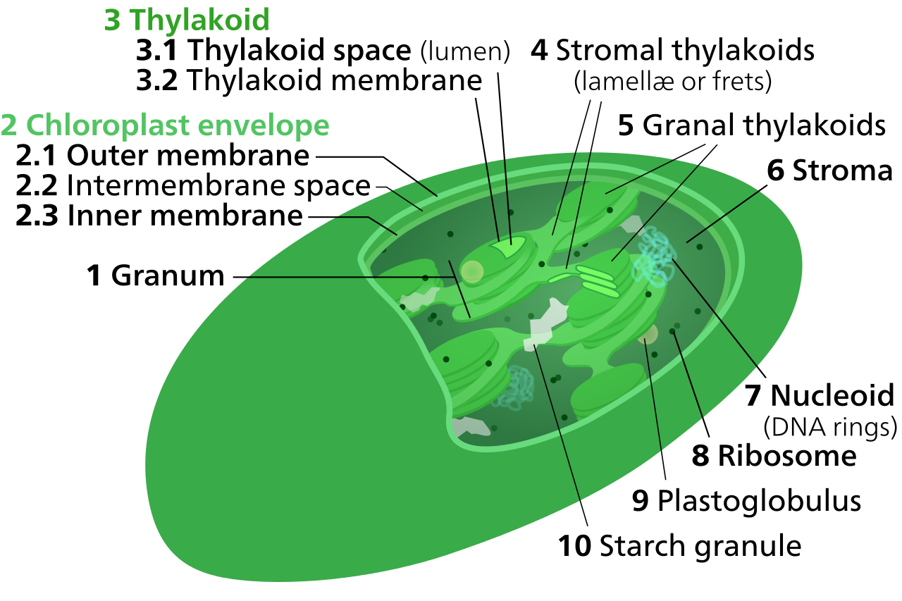
Parts of a chloroplast. (Source: Wikipedia-CC BY-SA 3.0)
Lenticels pores that act as stomata along stems and mature roots.
Mitochondria organelles carry out the process of cellular respiration.
Water can be transported through several physiological processes:
Capillary action: occurs due to the cohesion and adhesion properties of water that result in water molecules having attractions to each other (cohesion) and to the material in which the water is located (adhesion). This results in water rising up when it is in a narrow (capillary) tube. The same phenomenon occurs in plants too as water rises up the Xylem.
Root Pressure: Is the pressure created by the accumulation of minerals at the roots, which results in the movement of water into the root cells through osmosis and water is then forced up the Xylem.
Transpiration pull: The evaporation of water (transpiration) creates a pull on the adjacent water molecules.
Sieve cells are cells that are perforated and allow the movement of water between cells. Active transport moves sugars from palisade cells to the Phloem. From there, water flows through osmosis and distributes the sugars to cells that need these nutrients.
Stimuli is anything external to the plant that can influence a plant’s physiological process.
Phototropism is the growth of a plant in response to light stimulus (towards a light source).
Gravitropism is the growth of a plant in response to gravitation stimuli.
Positive tropisms: growth that occurs towards the stimulus.
Negative tropisms: growth that occurs away from the stimulus.
A hormone called auxin is synthesized in the tip then it diffused down the stem, where it stimulates cells to become elongated resulting in bending toward the light source.
Gravitropism is mediated mostly by starch molecules/particles as they are heavy and move to the bottom of the root tip cells allowing plants to detect which way is down.
You can access Tensai High School Biology revision questions HERE